Ahpra registration PHY0002298174
How these guides are written and reviewed →
The electrocardiogram (ECG) is the monitor physiotherapists watch most during exercise. It records the heart's electrical signals through electrodes on the chest and limbs. It shows how fast the heart is going, whether the rhythm is regular, and whether any part of the heart muscle is short of blood or has been damaged. It takes seconds, is painless, and is one of the most useful tests in medicine. Physiotherapists working in cardiac rehabilitation and hospital wards are often watching a monitor during exercise, so recognising a normal trace — and knowing which changes mean stop and get help — is a core safety skill. This page explains how to read one systematically.
What the trace represents
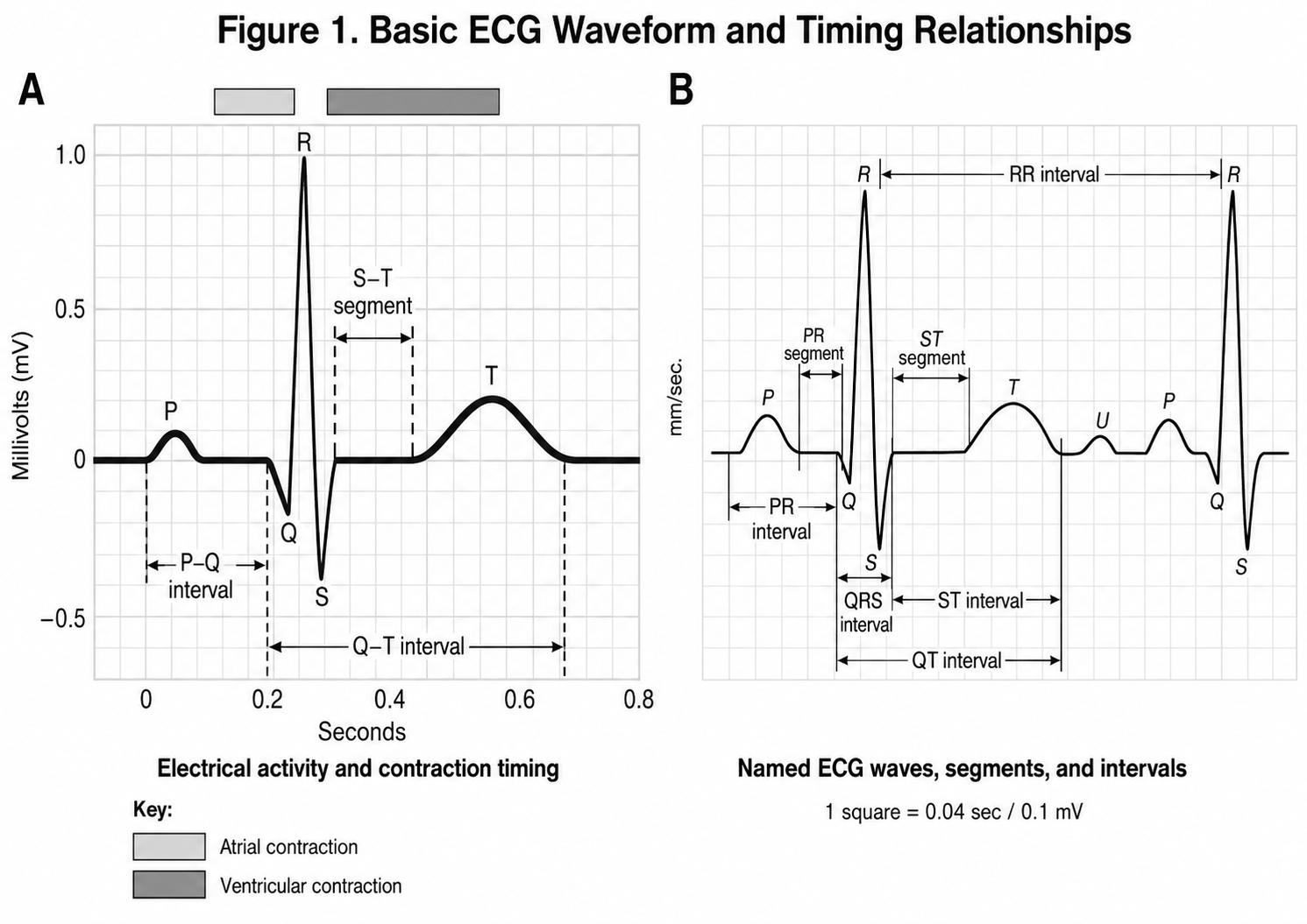
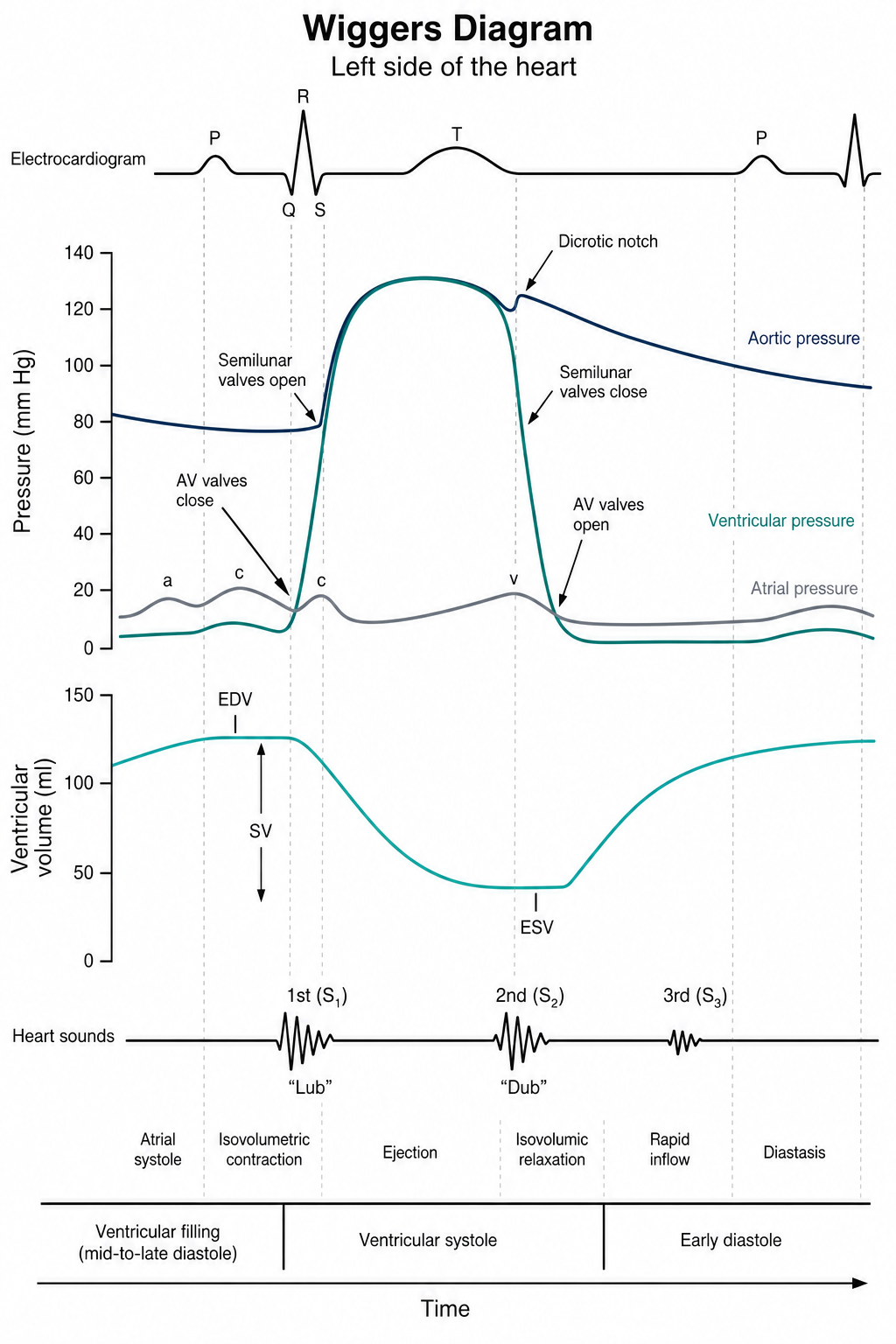
Each cardiac cycle produces a predictable sequence. The P wave is atrial depolarisation. The PR interval is the delay through the atrioventricular node. The QRS complex is ventricular depolarisation — the electrical event that produces the pulse. The ST segment is the period between depolarisation and repolarisation, and it is where ischaemia and infarction show. The T wave is ventricular repolarisation. A standard 12-lead ECG views this from twelve electrical angles; a single-lead or three-lead monitor shows rhythm only, and cannot exclude ischaemia.
Normal intervals at standard settings
Paper speed is 25 mm/s, so one small square is 0.04 s and one large square 0.20 s. Standard calibration is 10 mm per mV.
| Feature | Normal | Abnormal suggests |
|---|---|---|
| Rate | 60–100 bpm | Bradycardia or tachycardia — interpret against medication and context |
| P wave | Present, upright in II, one per QRS | Absent or chaotic: atrial fibrillation or flutter |
| PR interval | 120–200 ms (3–5 small squares) | Long: first-degree block. Progressive or dropped: higher-degree block |
| QRS duration | < 120 ms (< 3 small squares) | Wide: bundle branch block, ventricular origin, paced rhythm |
| ST segment | At the isoelectric line | Elevation: infarction or pericarditis. Depression: ischaemia or strain |
| QT interval | Corrected QT < 440 ms (men) / < 460 ms (women) | Prolonged: arrhythmia risk, often drug- or electrolyte-related |
A systematic reading
- Check the details. Right patient, right date and time, standard calibration and paper speed, and correct lead placement — limb-lead reversal is a common cause of an alarming-looking normal ECG.
- Rate. Divide 300 by the number of large squares between two R waves. For an irregular rhythm, count the complexes in a 6-second strip and multiply by ten.
- Rhythm. Regular or irregular? Is there a P wave before every QRS, and a QRS after every P?
- Axis. Broadly normal if leads I and II are both predominantly positive.
- Intervals. PR, QRS width, QT.
- Morphology and ST segments. Work through the leads in territories — inferior (II, III, aVF), lateral (I, aVL, V5, V6), anterior and septal (V1–V4) — looking for ST shift, T-wave inversion and pathological Q waves.
- Compare with the previous ECG. This is the single most valuable step. New change matters; long-standing change usually does not.
Rhythms a physiotherapist should recognise
| Rhythm | Recognition | What it means for the session |
|---|---|---|
| Sinus rhythm | P before every QRS, regular, 60–100 | Proceed |
| Sinus tachycardia | Same, > 100 | Look for a cause — pain, fever, anxiety, hypovolaemia, sepsis, embolism — before attributing it to effort |
| Atrial fibrillation | Irregularly irregular, no discernible P waves | Prescribe by RPE, not heart rate; see Atrial Fibrillation |
| Atrial flutter | Sawtooth baseline, often regular at ~150 | As for AF; a new fast flutter needs medical review |
| Ectopic beats (atrial or ventricular) | Early, often wide, complexes with a compensatory pause | Isolated ectopy is common and usually benign; increasing frequency with exercise, couplets or runs is not |
| Ventricular tachycardia | Broad, regular, fast (> 120), no P waves | Medical emergency — stop, assess responsiveness and pulse, call for help |
| Heart block | Long PR (first degree), dropped beats (second degree), P and QRS dissociated (third degree) | First degree is usually benign; second and third degree need medical assessment before exercise |
| Paced rhythm | Pacing spikes before P and/or QRS | Know the device, the programmed rate and the defibrillator therapy zone |
Ischaemia and infarction
Three patterns matter. ST elevation in two or more contiguous leads is acute infarction until proven otherwise — a time-critical emergency. ST depression and T-wave inversion suggest ischaemia or strain, and are what may appear during exercise testing in significant coronary disease. Pathological Q waves indicate established prior infarction and are old news unless new.
Territory maps to vessel: inferior leads (II, III, aVF) to the right coronary artery, anteroseptal (V1–V4) to the left anterior descending, lateral (I, aVL, V5–V6) to the circumflex. Knowing the territory of a previous infarct is useful context when assessing a patient in cardiac rehabilitation.
- New ST elevation, or new ST depression greater than 1–2 mm, particularly with symptoms.
- Ventricular tachycardia, or increasing ventricular ectopy with exercise — couplets, triplets or runs.
- New atrial fibrillation with a fast ventricular response, or any new arrhythmia with symptoms.
- New second- or third-degree heart block.
- Chest pain, syncope or pre-syncope, or a fall in systolic blood pressure with increasing workload.
- A defibrillator shock during a session — stop, sit the patient down, escalate, and do not resume that day.
Exercise testing and monitoring
Exercise ECG is used less for diagnosis than it once was, since imaging and CT coronary angiography perform better, but it remains useful for functional capacity, symptom reproduction, blood-pressure response and arrhythmia provocation.1 Standard termination criteria — ST depression beyond defined thresholds, sustained ventricular arrhythmia, a fall in systolic pressure, moderate-to-severe angina, or the patient's request — are published and should be known by anyone supervising a test.2
In cardiac rehabilitation, continuous ECG monitoring is not required for most low-risk patients; risk stratification determines who needs it, and over-monitoring adds cost and anxiety without benefit.3 Wearable and smartwatch single-lead ECGs now detect atrial fibrillation with reasonable accuracy and increasingly arrive with the patient, but they cannot exclude ischaemia and should prompt formal assessment rather than substitute for it.4
Common pitfalls
- Artefact is more common than pathology. Shivering, movement, poor electrode contact and electrical interference mimic arrhythmia — look at the patient before you look at the trace.
- The monitor is not a 12-lead. A three-lead monitor answers rhythm questions only.
- Heart rate is unreliable in atrial fibrillation: pulse deficit means the peripheral pulse and the ECG rate can differ substantially.
- Beta blockade blunts the rate response — a heart rate of 95 at peak effort may still be maximal for that patient.
- Automated interpretation is a prompt, not a diagnosis. Machine reads are frequently wrong, especially with artefact or paced rhythms.
- An ECG is one moment. A normal trace does not exclude intermittent arrhythmia or ischaemia; symptoms warrant referral regardless.
Scope of practice
Physiotherapists in Australia are not the primary interpreters of the ECG, and it is not a physiotherapy diagnosis. What is expected in cardiorespiratory and cardiac rehabilitation practice is safe recognition: identifying normal sinus rhythm, recognising the arrhythmias and changes that make exercise unsafe, understanding the patient's baseline trace and device settings before prescribing, and escalating clearly and early. Where local protocols permit physiotherapists to record a 12-lead ECG, technique and lead placement matter as much as reading — a poorly recorded trace generates false alarms and wastes clinical time.
What this changes in practice
In cardiac rehabilitation and acute cardiorespiratory work, the physiotherapist is frequently the clinician present when a rhythm changes under load. That makes ECG competence a safety skill rather than an academic one. It shapes the prescription — knowing to abandon heart-rate targets in atrial fibrillation, to train below a defibrillator's therapy zone, to interpret a blunted rate response on beta blockade. It shapes escalation, because recognising ventricular tachycardia or new ST change in the gym determines the outcome. And it shapes credibility in the multidisciplinary team: a physiotherapist who can describe what they saw, on which lead, at what workload, with what symptoms, is contributing clinical data rather than an impression.
Evidence summary
Framing. The physiotherapist's ECG competency is not diagnostic interpretation but safe recognition under load: identifying baseline rhythm and conduction, detecting exercise-induced change, applying published termination criteria, and escalating appropriately. This matters because exercise-based cardiac rehabilitation is delivered largely by physiotherapists and exercise physiologists, and the event rate — while very low — is not zero.2,3
Safety and risk stratification- The absolute risk of a cardiovascular event during supervised exercise is very low, with high-intensity interval training carrying risk comparable to moderate-intensity exercise in coronary populations — supporting individualised rather than uniformly capped prescription.5
- Continuous ECG monitoring is not required for low-risk patients in cardiac rehabilitation; guidance directs monitoring by risk stratification, and routine telemetry for all adds cost without demonstrated benefit.3
- Standardised exercise-testing termination criteria (ST depression thresholds, sustained ventricular arrhythmia, exertional hypotension, moderate-to-severe angina, neurological symptoms) are published and should be operationalised in local protocols.2
- Exercise ECG has modest diagnostic accuracy for obstructive coronary disease and has been displaced by imaging and CT coronary angiography in guideline diagnostic pathways — its remaining value is functional and prognostic.1
- Atrial fibrillation makes heart rate an unreliable prescription variable because of beat-to-beat variability and pulse deficit; use RPE, the talk test or a workload target, and treat wrist-worn optical readings as approximate.6
- Exercise-induced ventricular ectopy that increases in frequency or complexity with workload, and particularly ectopy in recovery, carries prognostic significance and warrants review rather than continued progression.7
- With an implantable defibrillator, train approximately 20 bpm below the programmed therapy zone, know the device settings before the first session, and have a documented plan for a shock during exercise.8
- Cardiac sarcoidosis, ARVC, hypertrophic cardiomyopathy and myocarditis each have specific arrhythmic risk profiles that override generic exercise rules — obtain the treating cardiologist's parameters.8
- Automated ECG interpretation is unreliable in the presence of artefact, paced rhythms and conduction abnormality, and should never be relied on without human review.9
- Smartwatch and single-lead consumer ECGs detect atrial fibrillation with reasonable sensitivity but generate false positives and cannot assess ischaemia; treat a patient-supplied trace as a prompt for formal assessment.4
- Lead misplacement — particularly limb-lead reversal and high V1–V2 placement — is a frequent source of pseudo-pathology and should be excluded before escalating an unexpected finding.9
- Review the baseline 12-lead and the device settings before the first exercise session in any cardiac patient — rhythm, conduction, prior infarct territory, pacing, therapy zone.
- Prescribe by RPE and symptoms wherever heart rate is unreliable: atrial fibrillation, beta blockade, pacing, chronotropic incompetence.
- Look at the patient before the monitor. Most alarms are artefact; the exceptions announce themselves clinically.
- Document specifically — rhythm, rate, lead, workload, symptoms, blood pressure and response to cessation — because that is what the medical team needs to act on.
- Know your local escalation pathway and resuscitation equipment location before you need them, and rehearse the defibrillator-shock scenario with the team.
- New change is what matters; compare with the previous trace before treating an abnormality as acute.
- A blunted heart-rate response is expected on beta blockade and is not chronotropic incompetence unless the trace and the effort say so.
- Symptoms with a normal trace still warrant referral — intermittent arrhythmia and ischaemia are common and a single ECG is one moment in time.
- Sinus tachycardia is a symptom, not a diagnosis: find the cause before attributing it to deconditioning.
- No study has evaluated whether physiotherapist ECG competency affects adverse-event detection or patient outcomes in rehabilitation.
- Optimal monitoring intensity for intermediate-risk cardiac rehabilitation participants is not established.3
- Thresholds for stopping exercise on ventricular ectopy are consensus-based rather than trialled.7
- How consumer wearable ECG data should be integrated into rehabilitation decision-making is undefined.4
References & evidence base
- Vrints C, Andreotti F, Koskinas KC, et al. 2024 ESC guidelines for the management of chronic coronary syndromes. Eur Heart J 2024;45(36):3415–3537.
- American College of Sports Medicine. ACSM's guidelines for exercise testing and prescription. 11th ed. Philadelphia: Wolters Kluwer; 2021.
- Woodruffe S, Neubeck L, Clark RA, et al. Australian Cardiovascular Health and Rehabilitation Association (ACRA) core components of cardiovascular disease secondary prevention and cardiac rehabilitation 2014. Heart Lung Circ 2015;24(5):430–441.
- Perez MV, Mahaffey KW, Hedlin H, et al. Large-scale assessment of a smartwatch to identify atrial fibrillation. N Engl J Med 2019;381(20):1909–1917.
- Rognmo Ø, Moholdt T, Bakken H, et al. Cardiovascular risk of high- versus moderate-intensity aerobic exercise in coronary heart disease patients. Circulation 2012;126(12):1436–1440.
- Van Gelder IC, Rienstra M, Bunting KV, et al. 2024 ESC guidelines for the management of atrial fibrillation. Eur Heart J 2024;45(36):3314–3414.
- Frolkis JP, Pothier CE, Blackstone EH, Lauer MS. Frequent ventricular ectopy after exercise as a predictor of death. N Engl J Med 2003;348(9):781–790.
- Pelliccia A, Sharma S, Gati S, et al. 2020 ESC guidelines on sports cardiology and exercise in patients with cardiovascular disease. Eur Heart J 2021;42(1):17–96.
- Kligfield P, Gettes LS, Bailey JJ, et al. Recommendations for the standardization and interpretation of the electrocardiogram: part I. Circulation 2007;115(10):1306–1324.
References are numbered in citation order (Vancouver/BMJ style) and were current at the time of writing. Guidelines are living documents — verify against the latest version before clinical use.
Corrections: If something on this page is wrong, out of date or unclear, we want to know. Email reception@inspireclinic.au with the page name and what you believe is incorrect. Substantive corrections are made promptly, and the guide’s version and last-updated date are changed to reflect it.