Ahpra registration PHY0002298174
How these guides are written and reviewed →
Oxygen is a drug. It is prescribed to a target range of oxygen saturation, given through a device chosen to match the patient, then titrated and weaned like any other medicine — not left running at a fixed rate. Too little is dangerous; in a specific group of patients, too much is also dangerous.
Oxygen is the most commonly given drug in acute care and one of the least often prescribed properly. It treats hypoxaemia — a low level of oxygen in the arterial blood — and nothing else. It does not treat breathlessness in a patient who is not hypoxaemic, and it does not treat the underlying cause of the hypoxaemia.1 The clinical decisions are therefore always the same three: what saturation am I aiming for, which device delivers it, and when do I come down.
Oxygen is a prescribed drug
Oxygen should be prescribed to a target saturation range rather than a fixed flow rate, with the device and starting flow recorded, and then adjusted by the person delivering care to keep the patient inside the range.1 The evidence for restraint is now strong: a large systematic review and meta-analysis of acutely ill adults found that liberal oxygen increased mortality with no compensating benefit, which drove the guidance away from routine high-concentration oxygen for everyone.2,3
Target saturation ranges
| Patient group | Target SpO2 | Notes |
|---|---|---|
| Most acutely unwell adults | 92–96% | The Australian target range, set by the Thoracic Society of Australia and New Zealand (TSANZ) and the Australian and New Zealand Committee on Resuscitation (ANZCOR).4 Do not aim higher — above 96% there is no benefit and possible harm in several conditions. British guidance (BTS) uses 94–98%, so a range quoted from a UK source will sit two points higher1 |
| At risk of hypercapnic respiratory failure | 88–92% | chronic obstructive pulmonary disease (COPD), obesity hypoventilation, neuromuscular and chest-wall disease, bronchiectasis, cystic fibrosis, known CO2 retention. Use a controlled-delivery device |
| Critical illness / resuscitation | Highest available initially | Reservoir mask at 15 L/min while stabilising, then titrate down as soon as saturation and blood gases allow |
| Not hypoxaemic | No oxygen | Breathlessness without hypoxaemia is not an indication. Treat the cause; consider a handheld fan and breathing control |
A patient at risk of hypercapnic failure should carry that information with them — an alert card, a documented target range, and ideally a previous blood gas. The 88–92% target is a starting point until a gas is available, not a permanent ceiling.1
Why high-concentration oxygen raises carbon dioxide
The old teaching — that oxygen removes the hypoxic drive to breathe and the patient simply stops breathing — is a small part of a larger mechanism and on its own is misleading. The dominant contributors are release of hypoxic pulmonary vasoconstriction, which worsens ventilation–perfusion matching and increases dead-space ventilation, and the Haldane effect, whereby oxygenated haemoglobin releases carbon dioxide it was carrying. Reduced respiratory drive contributes but is rarely the main event.5
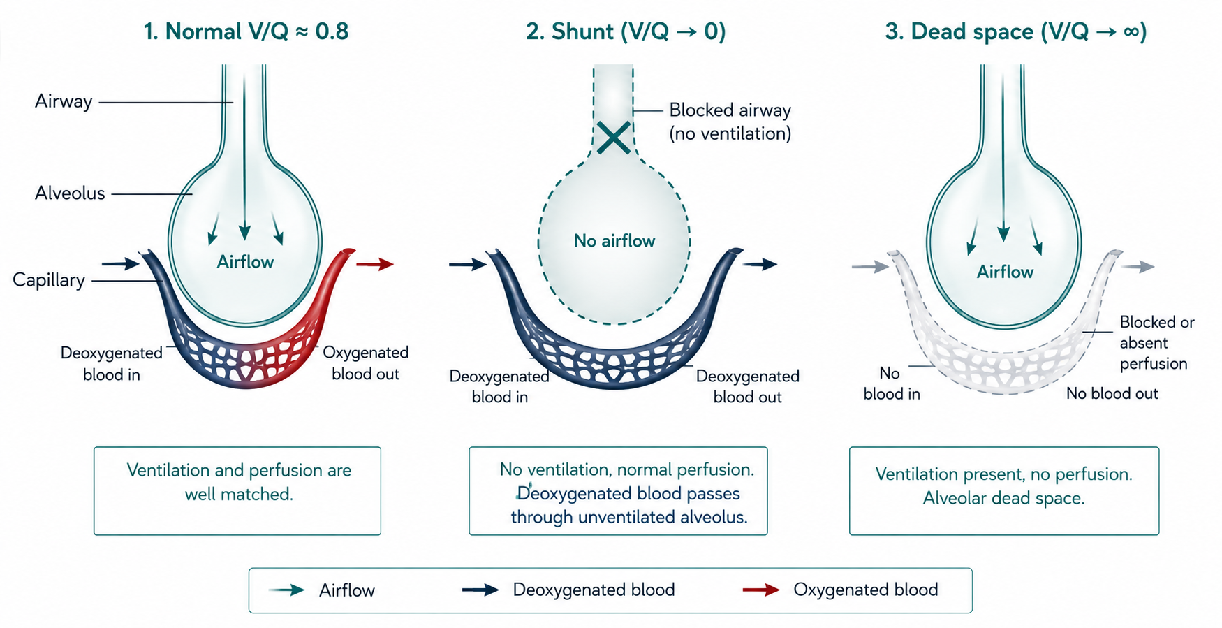
Shunt — blood passes lung that is perfused but not ventilated, so it returns to the systemic circulation still deoxygenated. Shunt responds poorly to added oxygen, which is why a large shunt produces hypoxaemia that is stubborn to supplemental oxygen and why recruitment, positioning and clearance matter more than turning the flow up.
Dead space — lung that is ventilated but not perfused. It does not cause hypoxaemia directly but wastes each breath, raising the work of breathing and impairing carbon dioxide clearance.
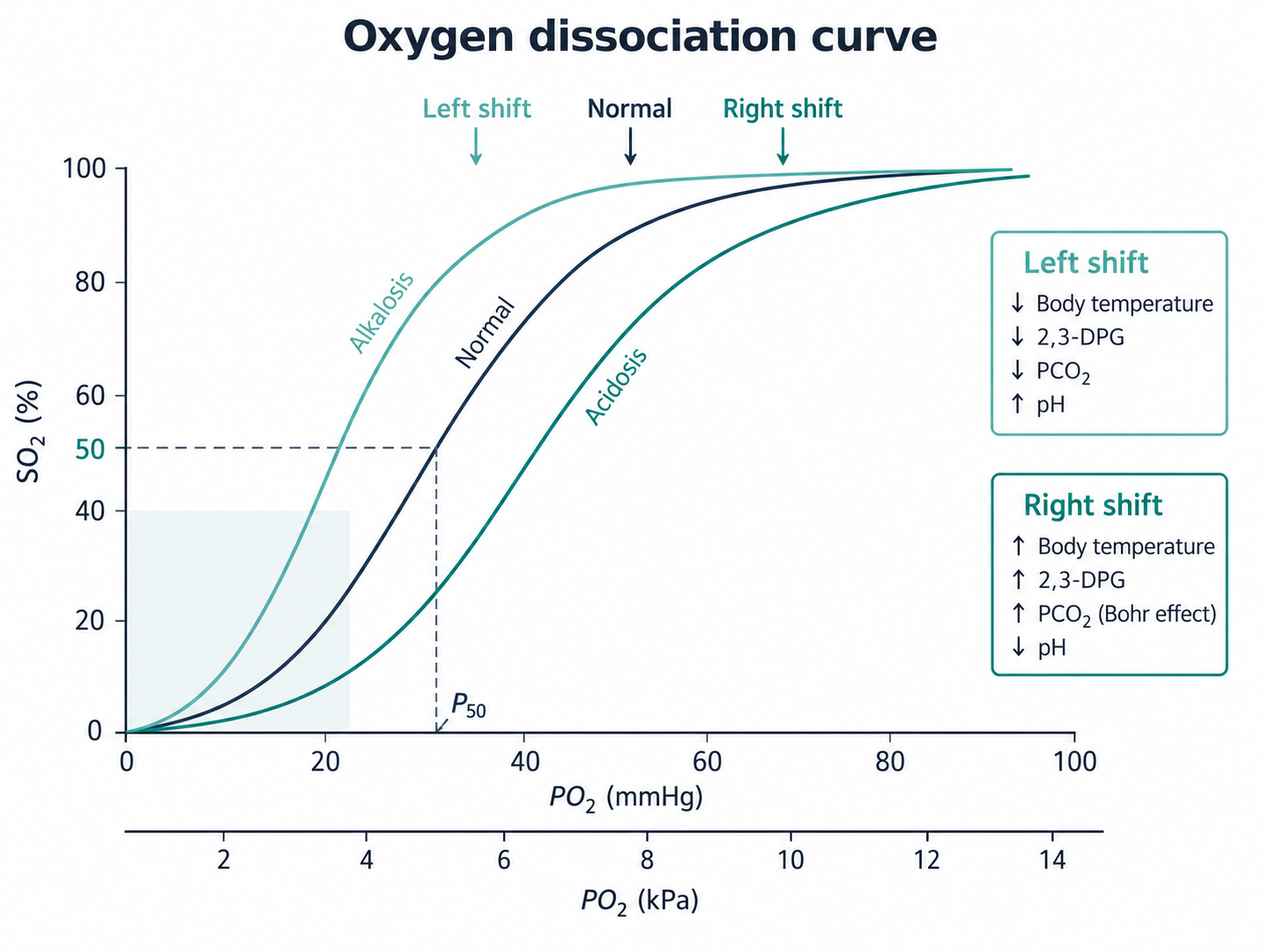
The sigmoid shape is the clinically important part. Above roughly 90% saturation the curve is flat, so large changes in arterial oxygen tension produce almost no change in saturation — a patient can be losing considerable reserve while the oximeter barely moves.
Below about 90% the curve is steep: a small further fall in tension causes a rapid fall in saturation. This is why a patient who begins to desaturate during exercise or treatment can deteriorate quickly, and why 88–92% is a range that needs watching rather than a comfortable place to sit.
Low-flow (variable, uncontrolled) delivery
With a low-flow device, the delivered flow is less than the patient’s peak inspiratory flow rate, so they entrain room air to make up the difference. The delivered concentration therefore varies breath to breath with their respiratory pattern — a patient breathing hard entrains more air and receives a lower concentration than the same device on a settled patient. Low-flow devices are comfortable and convenient, but they are the wrong choice when the concentration must be known.1
Nasal cannula
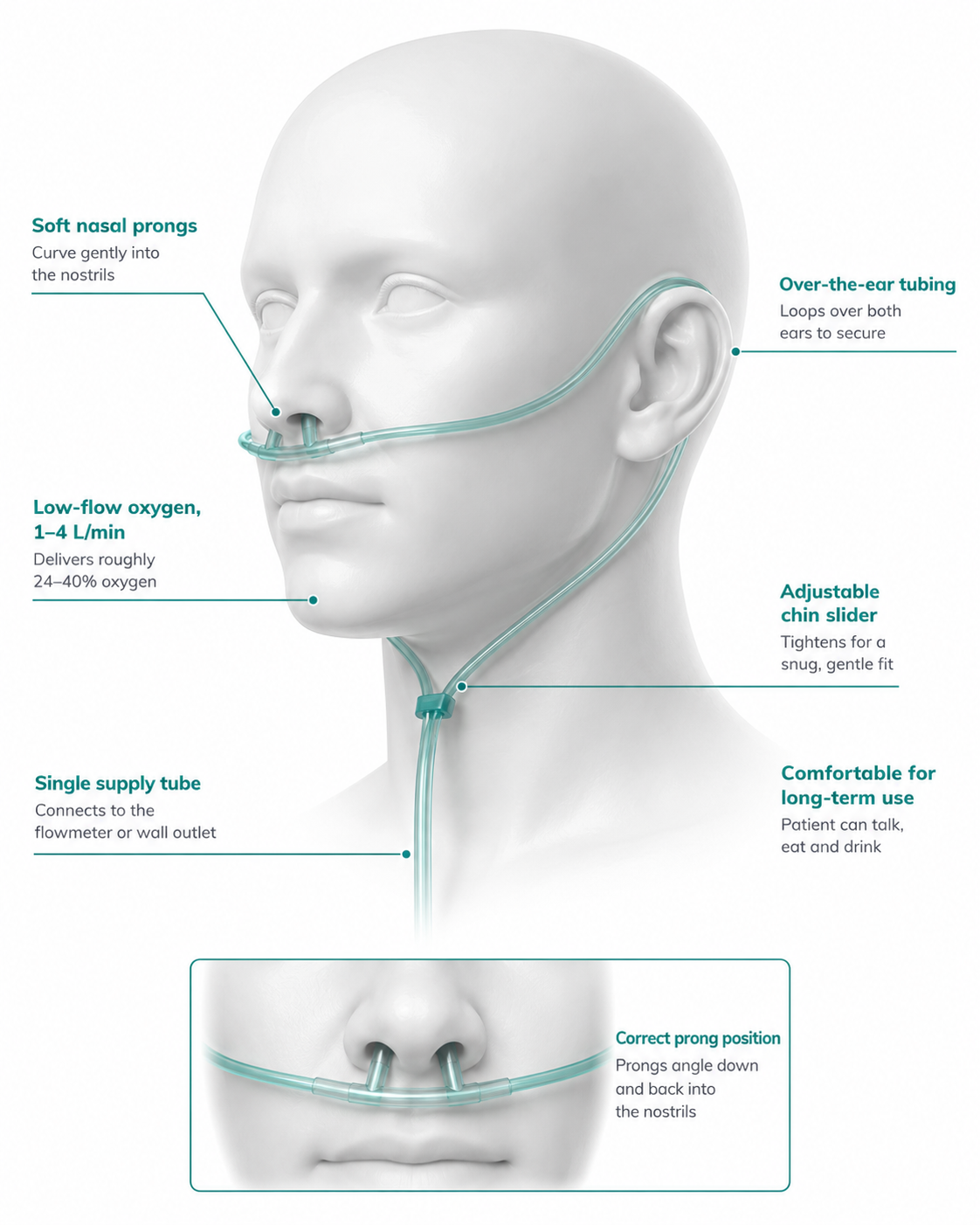
1–4 L/min, giving roughly 24–40% oxygen. Above 4 L/min it becomes uncomfortable and drying without adding much.
The practical advantage is that the patient can eat, drink and talk, which makes it the device of choice for mobilising, for rehabilitation and for anyone staying on oxygen for days. Check the ears and nostrils for pressure damage from the tubing.
Simple (Hudson) mask
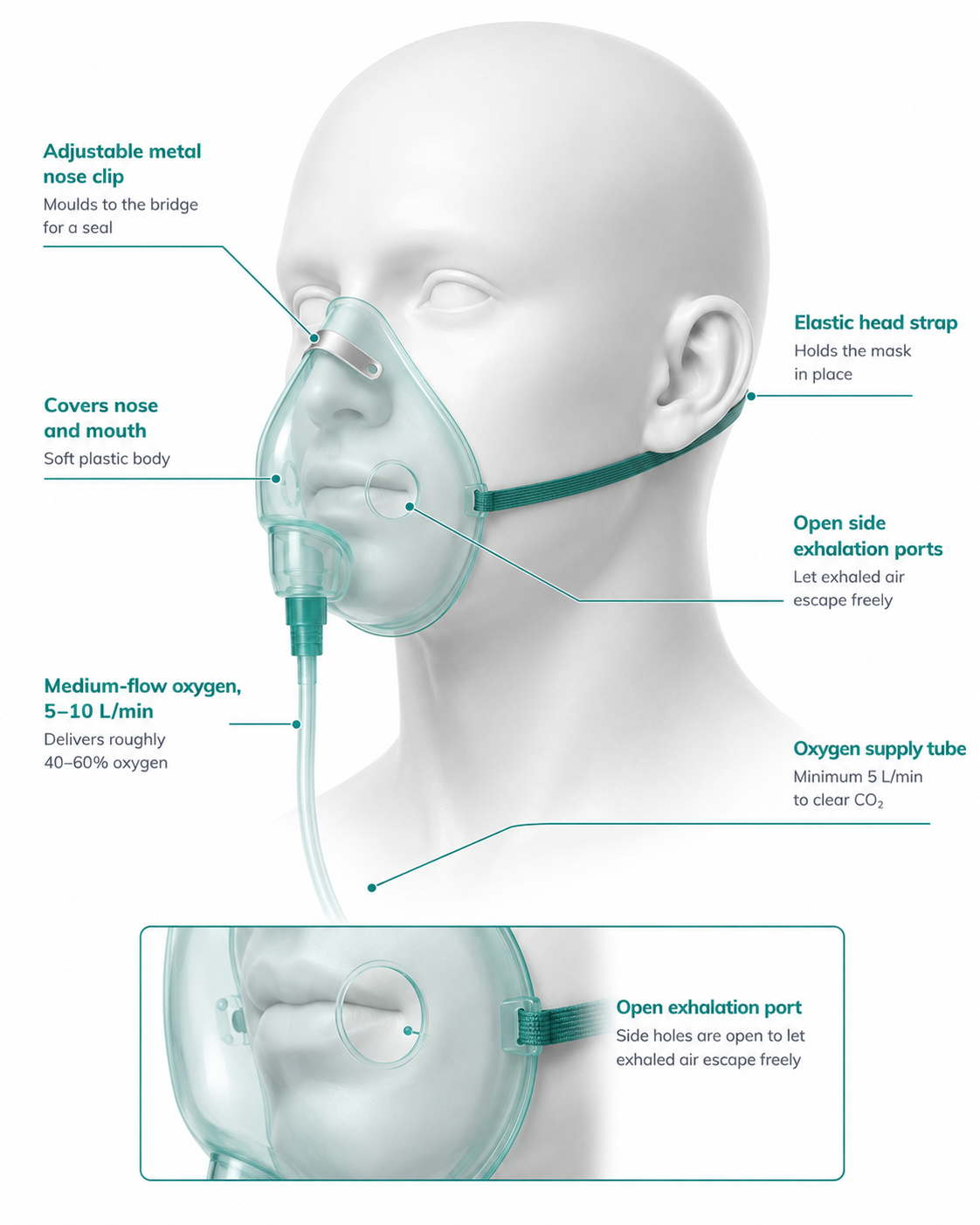
5–10 L/min, giving roughly 40–60% oxygen. It must not be run below 5 L/min — at low flows exhaled gas is not flushed from the mask and the patient rebreathes their own carbon dioxide.
Interferes with eating and speech, and the delivered concentration is unpredictable. Rarely the right long-term choice; a Venturi mask does the same job with a known concentration.
Reservoir (non-rebreathe) mask
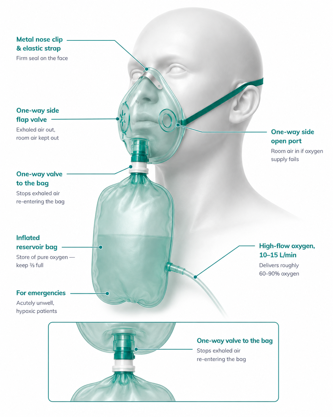
15 L/min, giving roughly 60–90% oxygen. Inflate the reservoir bag before applying it and keep the flow high enough that the bag does not collapse on inspiration.
This is an emergency and stabilisation device, not a maintenance one. A patient still on a reservoir mask after the first hours needs review, a blood gas and a plan — not another shift on 15 L/min.
Fixed-performance delivery — the Venturi mask
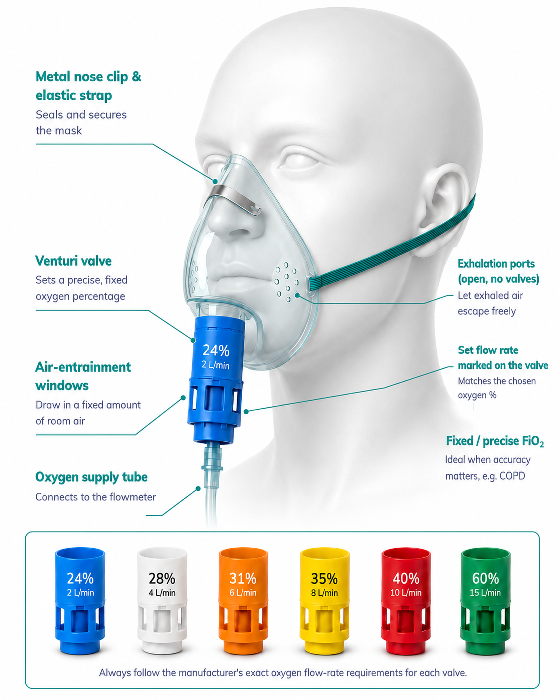
A Venturi valve entrains a fixed ratio of room air to oxygen, delivering a flow that exceeds the patient’s peak inspiratory flow. The concentration is therefore known and stable regardless of how the patient is breathing.
Each colour-coded valve states its concentration and the minimum oxygen flow needed to achieve it — run it at or above that flow or the delivery is no longer fixed. In a breathless patient, increase the driving flow above the stated minimum; the concentration stays the same.
This is the device for a patient at risk of hypercapnic respiratory failure, where an unknown concentration is precisely the problem: 24% and 28% valves allow controlled oxygen while a blood gas is obtained and reviewed.1
High-flow nasal oxygen (HFNO)
high-flow nasal oxygen delivers heated, humidified gas at up to about 60 L/min with the concentration set independently of flow, up to approximately 100%. Because the flow exceeds the patient’s inspiratory demand, entrainment of room air is reduced and the delivered concentration is reliable. Additional mechanisms are washout of nasopharyngeal dead space, a small positive airway pressure effect, and better secretion clearance from full humidification.6,7
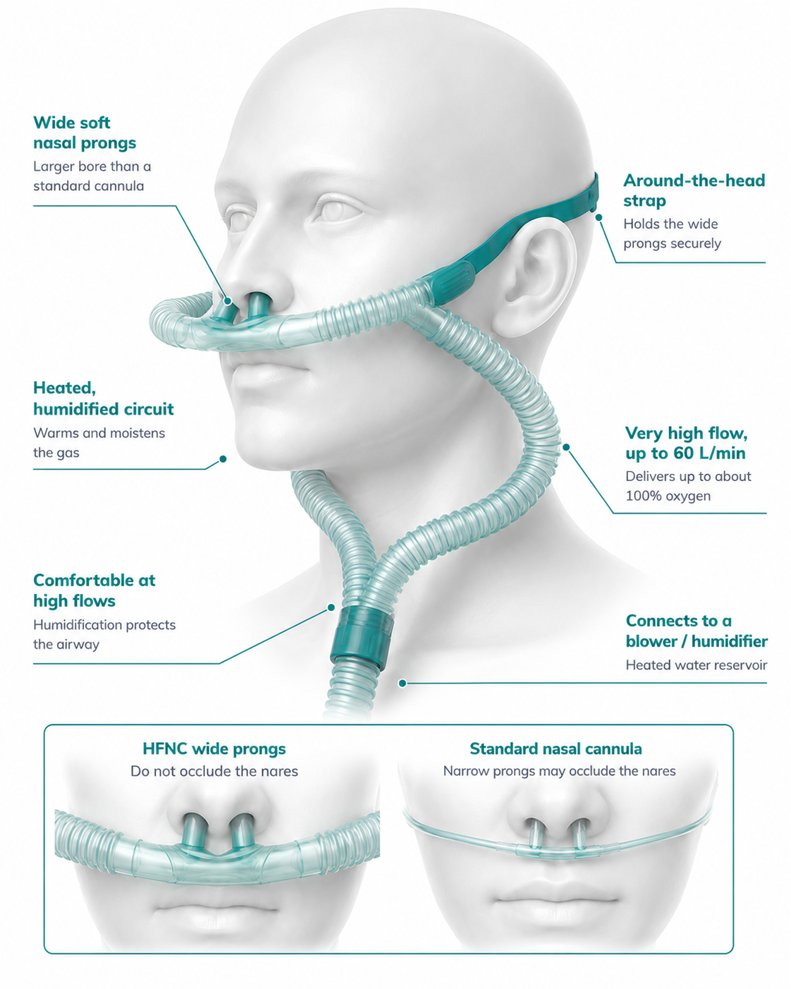
The wide, soft prongs are deliberately sized not to occlude the nares — gas must be able to escape around them, or pressure builds. This is the main set-up error to look for.
Full humidification is what makes high flows tolerable and protects the mucosa. It also means the circuit runs warm and wet: check for condensate, and keep the water chamber below the patient.
Indications, evidence and failure
In acute hypoxaemic respiratory failure, high-flow nasal oxygen reduced escalation to invasive ventilation compared with conventional oxygen and non-invasive ventilation in FLORALI, with a mortality signal in the most hypoxaemic subgroup.6 European guidance suggests high-flow over conventional oxygen in acute hypoxaemic failure, and after extubation in patients at risk of reintubation.7 It is not a treatment for hypercapnic failure, where non-invasive ventilation remains the intervention with the evidence.
The danger of high-flow is that it makes a deteriorating patient look comfortable while the underlying failure progresses. The ROX index — saturation divided by inspired oxygen concentration, divided by respiratory rate — was developed to identify this: a low or falling value predicts high-flow failure and should prompt escalation rather than another increase in flow.8 Rising respiratory rate, accessory muscle use, falling ROX and rising carbon dioxide are the signs that matter.
Humidification
Normal airway conditioning warms inspired gas to body temperature and fully saturates it with water by the time it reaches the carina. Dry medical gas at high flow, or any flow bypassing the upper airway through a tracheostomy or endotracheal tube, defeats this — drying secretions, impairing ciliary function and making sputum harder to clear.
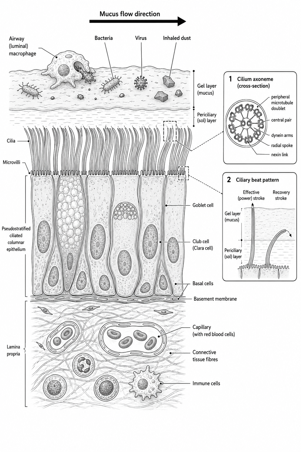
Cilia beat within a thin layer of watery periciliary fluid beneath the mucus blanket. If that layer dries or thickens, the cilia cannot beat effectively and the escalator stalls — secretions become tenacious and retained.
Humidify for any patient with an artificial airway, on high-flow, needing oxygen beyond a day or two, or whose secretions are thick and difficult to clear. Warmed humidification is the most effective; a cold bubble humidifier adds little.
Oxygen in specific situations
- Acute coronary syndrome. Routine oxygen in patients who are not hypoxaemic is not beneficial and was associated with larger infarct size in the AVOID trial. Give oxygen only for hypoxaemia.9
- Stroke. No routine oxygen for non-hypoxaemic patients; treat hypoxaemia to the usual target.1
- Cardiac arrest. Highest available concentration during resuscitation, then titrate promptly after return of spontaneous circulation — sustained hyperoxaemia after arrest is harmful.1
- Palliative breathlessness. In patients who are breathless but not hypoxaemic, oxygen is no better than medical air; a handheld fan, positioning, breathing control and opioids are the interventions with evidence.10
Long-term and ambulatory oxygen
Long-term oxygen therapy improves survival in chronic, stable hypoxaemia and must be assessed on stable-state arterial blood gases, not during or shortly after an exacerbation — hypoxaemia during an acute event frequently resolves. Australian and British guidance both require repeat measurement at least several weeks after recovery, and both set thresholds around a resting arterial oxygen tension at or below 55 mmHg, or below 60 mmHg with evidence of end-organ effect such as polycythaemia, pulmonary hypertension or cor pulmonale.11,12
Two points that are regularly got wrong: long-term oxygen requires at least 15 hours a day to deliver the survival benefit, and it is not a treatment for breathlessness. Prescribing it to a patient who does not meet the gas criteria adds burden, cost and a fire risk without benefit. Smoking is an absolute contraindication.11,12
Safety
Oxygen is an oxidising agent and vigorously supports combustion. No smoking, no naked flame, no emollients containing paraffin near the face, and no oil or grease on fittings. Cylinders are stored upright and secured. In the home, working smoke alarms and a route out matter as much as the prescription.11
Implications for physiotherapy
- Read the prescription before you treat. Know the target range and whether the patient is at risk of hypercapnic failure. A patient on 88–92% is a different treatment proposition from one on 92–96%.
- Record the device and flow with every saturation. “SpO2 92%” means nothing on its own; 92% on 1 L/min and 92% on 15 L/min are different patients.
- Titrate during exercise, then wean afterwards. Many patients need more oxygen to walk than to sit. Increase to hold the target during the session and bring it back down afterwards — and hand the flow back to the nursing team explicitly.
- Desaturation on exertion is an assessment finding. Note the nadir, the flow required, and the recovery time. Because of the shape of the dissociation curve, a fall below 90% can accelerate.
- Positioning and clearance often do more than flow. Where hypoxaemia is driven by shunt — consolidation, atelectasis, retained secretions — recruitment and clearance address the cause; more oxygen only treats the number.
- Swap the device to suit the treatment. A patient in a mask who needs to mobilise and talk is usually better served by a cannula at an equivalent target, if the concentration allows.
References & evidence base
- O’Driscoll BR, Howard LS, Earis J, Mak V. BTS guideline for oxygen use in adults in healthcare and emergency settings. Thorax 2017;72(Suppl 1):ii1–ii90.
- Siemieniuk RAC, Chu DK, Kim LH, et al. Oxygen therapy for acutely ill medical patients: a clinical practice guideline. BMJ 2018;363:k4169.
- Chu DK, Kim LH, Young PJ, et al. Mortality and morbidity in acutely ill adults treated with liberal versus conservative oxygen therapy (IOTA): a systematic review and meta-analysis. Lancet 2018;391(10131):1693–705.
- Barnett A, Beasley R, Buchan C, Chien J, Farah CS, King G, et al. Thoracic Society of Australia and New Zealand Position Statement on Acute Oxygen Use in Adults: ‘Swimming between the flags’. Respirology 2022;27(4):262–276.
- Abdo WF, Heunks LMA. Oxygen-induced hypercapnia in COPD: myths and facts. Crit Care 2012;16(5):323.
- Frat J-P, Thille AW, Mercat A, et al. High-flow oxygen through nasal cannula in acute hypoxemic respiratory failure (FLORALI). N Engl J Med 2015;372(23):2185–96.
- Rochwerg B, Einav S, Chaudhuri D, et al. The role of high flow nasal cannula in acute respiratory failure: ERS clinical practice guidelines. Eur Respir J 2022;59(4):2101574.
- Roca O, Caralt B, Messika J, et al. An index combining respiratory rate and oxygenation to predict outcome of nasal high-flow therapy (ROX index). Am J Respir Crit Care Med 2019;199(11):1368–76.
- Stub D, Smith K, Bernard S, et al. Air versus oxygen in ST-segment-elevation myocardial infarction (AVOID). Circulation 2015;131(24):2143–50.
- Ekström M, Ahmadi Z, Bornefalk-Hermansson A, Abernethy A, Currow D. Oxygen for breathlessness in patients with chronic obstructive pulmonary disease who do not qualify for home oxygen therapy. Cochrane Database Syst Rev 2016;(11):CD006429.
- Hardinge M, Annandale J, Bourne S, et al. BTS guidelines for home oxygen use in adults. Thorax 2015;70(Suppl 1):i1–i43.
- McDonald CF, Whyte K, Jenkins S, Serginson J, Frith P. Clinical practice guideline on adult domiciliary oxygen therapy: Thoracic Society of Australia and New Zealand. Respirology 2016;21(1):76–8.
References are numbered in citation order (Vancouver/BMJ style) and were current at the time of writing. Guidelines are living documents — verify against the latest version before clinical use.
Setting up and reviewing home oxygen and non-invasive ventilation is a physiotherapy service in its own right, and the practical side of this is a large part of it.
Corrections: If something on this page is wrong, out of date or unclear, we want to know. Email reception@inspireclinic.au with the page name and what you believe is incorrect. Substantive corrections are made promptly, and the guide’s version and last-updated date are changed to reflect it.