Ahpra registration PHY0002298174
How these guides are written and reviewed →
In short
- Call 000 now: Chest pain, pressure or heaviness that lasts more than ten minutes, is not relieved by two doses of glyceryl trinitrate (GTN) five minutes apart, or comes with sweating, nausea, breathlessness or pain spreading to the arm, jaw or back.
- An exercise test before you start helps set the right level of exercise for you.
- Cardiac rehabilitation, led by physiotherapists, improves survival, symptoms and confidence after a heart event.
Coronary artery disease is narrowing of the arteries that supply the heart muscle, caused by a gradual build-up of fatty plaque. It can cause chest tightness or breathlessness on exertion (angina) and, if an artery blocks suddenly, a heart attack. Treatment combines lifestyle changes, medicines and sometimes procedures to open or bypass the narrowed arteries. Cardiac rehabilitation — supervised exercise and education — is one of the most effective ways to recover and prevent future problems. This page explains coronary artery disease and recovery.
Stable coronary artery disease — now termed "chronic coronary syndrome" (CCS) in international guidelines to emphasise its progressive nature — describes the spectrum of presentations between acute coronary events. It is the substrate from which most myocardial infarctions arise and a major focus of secondary prevention and cardiac rehabilitation.
Definition
Chronic coronary syndrome encompasses several clinical presentations: stable angina, ischaemia without obstructive coronary disease (INOCA), patients > 1 year following ACS or revascularisation, ischaemic cardiomyopathy, and vasospastic and microvascular angina. Each shares an underlying coronary atherosclerotic or microvascular substrate but differs in management focus.
Pathophysiology
Atherosclerotic plaque develops over decades, driven by endothelial dysfunction, lipid deposition, inflammation, smooth muscle proliferation, and (in advanced lesions) calcification. Symptomatic ischaemia occurs when oxygen demand exceeds supply, classically during exertion. Plaque rupture or erosion underlies acute coronary syndromes.
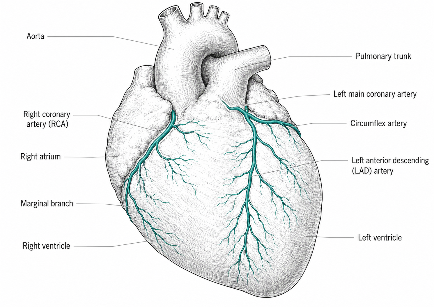
Microvascular angina reflects disease of the small coronary arterioles, particularly in women, and frequently coexists with typical angina or occurs in isolation.
Co-morbidities
Hypertension, diabetes, dyslipidaemia, obesity, chronic kidney disease, obstructive sleep apnoea (OSA), atrial fibrillation, peripheral arterial disease, and depression. The pattern of clustering largely defines individual risk trajectory.
Prevalence
Ischaemic heart disease remains the leading cause of death in Australia. Stable angina affects approximately 3–4% of adults, with rising prevalence with age.
Causes and risk factors
Modifiable: smoking, hypertension, dyslipidaemia, diabetes, obesity, physical inactivity, poor diet, excessive alcohol, and psychosocial stress.
Non-modifiable: increasing age, male sex (and post-menopausal female), family history of premature CVD, Aboriginal and Torres Strait Islander identity, South Asian ancestry, and inherited dyslipidaemias including familial hypercholesterolaemia.
Symptoms
Classic stable angina: retrosternal chest discomfort precipitated by exertion or emotion, relieved by rest or sublingual nitrate, lasting minutes. Atypical presentations are common in women, older adults, and those with diabetes (breathlessness, fatigue, jaw or arm discomfort).
Microvascular and vasospastic angina may present with rest pain, prolonged episodes, or atypical features.
Diagnosis
Why diagnosis matters
Establishing the diagnosis allows initiation of disease-modifying therapy (antiplatelet, statin, ACEi/ARB where indicated), assessment of revascularisation need, structured cardiac rehabilitation referral, and identification of patients at increased risk of future acute events.1
Pre-test probability and initial assessment
Diagnostic pathway is guided by pre-test probability of obstructive CAD based on age, sex, and symptom character (typical, atypical, non-anginal). 12-lead ECG, fasting lipids and glucose/HbA1c, FBC, U&E, TSH, and assessment of left ventricular function (echocardiography) are routine.
Functional and anatomical testing
Functional testing (exercise ECG, stress echocardiography, stress cardiac magnetic resonance imaging (MRI), myocardial perfusion imaging) detects ischaemia. CT coronary angiography is increasingly first-line in low-to-intermediate risk patients, with calcium scoring as an adjunct to risk stratification.
Invasive coronary angiography is reserved for those with high-risk features, ongoing symptoms despite optimal medical therapy, or where revascularisation is being considered.
INOCA and microvascular disease
Patients with angina and non-obstructive coronary disease at angiography warrant consideration of microvascular and vasospastic disease, increasingly assessed with invasive coronary function testing or non-invasive coronary flow reserve measurement.
Management
Treatment goals
Twin goals are symptom control (anti-anginal therapy, revascularisation where indicated) and event prevention (antiplatelet, statin, blood pressure control, diabetes optimisation, smoking cessation, structured exercise). The latter delivers most of the prognostic benefit.
Lifestyle and cardiac rehabilitation
Cardiac rehabilitation is recommended for all patients with stable CAD, with evidence for reduced cardiovascular mortality, hospital re-admission, and improved quality of life. Components include supervised exercise training (typically 8–12 weeks, twice-weekly), education on disease and risk factor modification, dietary counselling, smoking cessation support, psychological screening, and medication optimisation.2,3
For patients in regional and remote Central Queensland, home-based cardiac rehabilitation with telehealth supervision has comparable outcomes and is increasingly favoured. At Inspire Clinic, individualised and small-group programmes are offered alongside home-based delivery.
Anti-anginal therapy
First-line: beta-blocker or calcium channel blocker (or both if needed), titrated to symptoms and heart rate (resting 55–60 bpm target). Sublingual nitrate for acute episodes. Second-line agents include long-acting nitrates, ivabradine (in sinus rhythm with HR > 70 on beta-blocker), nicorandil, and ranolazine.
Disease-modifying therapy
Antiplatelet therapy (aspirin 100 mg daily; clopidogrel for aspirin-intolerant). High-intensity statin titrated to LDL-C < 1.8 mmol/L (and < 1.4 mmol/L in very high-risk patients per current ESC and NHFA-aligned targets). ACE inhibitor or ARB for patients with hypertension, diabetes, CKD, or LV dysfunction. SGLT2 inhibitor for patients with diabetes, heart failure, or CKD per current indications.
Revascularisation
Reserved for symptoms refractory to optimal medical therapy, or for prognostic benefit in selected anatomies (left main, three-vessel disease with reduced LV function). Choice between percutaneous coronary intervention (PCI) and coronary artery bypass grafting (CABG) is determined by anatomy, comorbidity, and patient preference, with multidisciplinary heart team discussion in complex cases.4,5,6
Identifying an exacerbation or acute event
New, prolonged, or rest-onset chest pain, or pain unresponsive to two doses of sublingual nitrate at five-minute intervals, mandates emergency presentation. Patients should be given a written action plan that specifies these criteria.
Medications
Antiplatelet therapy
Aspirin 100 mg daily indefinitely (clopidogrel 75 mg daily if aspirin-intolerant). Dual antiplatelet therapy following PCI for defined durations is managed by cardiology.
Statin and lipid management
High-intensity statin (atorvastatin 40–80 mg, rosuvastatin 20–40 mg). Ezetimibe added if LDL-C target not reached. PCSK9 inhibitors (evolocumab, alirocumab) and (under specialist criteria) inclisiran for high-risk patients not at target on maximally tolerated statin/ezetimibe.
Anti-anginal
Beta-blockers (bisoprolol, metoprolol, atenolol), non-dihydropyridine CCB (diltiazem, verapamil) or dihydropyridine CCB (amlodipine), long-acting nitrates (isosorbide mononitrate), ivabradine, nicorandil, and ranolazine as outlined above.
Renin-angiotensin and SGLT2 inhibitors
ACE inhibitors or ARBs in patients with hypertension, diabetes, CKD, or LV dysfunction. SGLT2 inhibitors (empagliflozin, dapagliflozin) increasingly used across CAD with diabetes, heart failure, or CKD given clear cardiovascular and renal benefit.
Multi-system manifestations
Concurrent vascular disease
Polyvascular disease (concurrent coronary, cerebrovascular, and peripheral arterial disease) is common. Patients with stable CAD warrant screening for cerebrovascular and peripheral disease where clinical features suggest, with attention to ankle-brachial index and carotid auscultation.
Heart failure
Ischaemic cardiomyopathy is the leading cause of heart failure. Patients with stable CAD and reduced LV function warrant heart failure-specific management in parallel.
Diabetes and metabolic disease
Tight glycaemic control without hypoglycaemia, blood pressure optimisation, and use of agents with proven cardiovascular benefit (SGLT2 inhibitors, GLP-1 receptor agonists in selected patients) form a core part of management.
Warning signs
Living with stable CAD
Exercise prescription
Following risk stratification, most patients with stable CAD can and should exercise regularly. Prescription combines aerobic activity (target heart rate set below the ischaemic threshold), resistance training (avoiding heavy isometric loading early), and warm-up and cool-down to minimise rebound symptoms. Symptom-limited exercise testing informs prescription.7,8
Sexual activity
Patients commonly fear cardiac events with sexual activity but the absolute risk is low in stable disease. Resumption is generally appropriate once moderate exertion is tolerated without symptoms. Sublingual nitrates are contraindicated within 24 hours (sildenafil, vardenafil) or 48 hours (tadalafil) of phosphodiesterase-5 inhibitor use.
Driving and occupational considerations
Austroads Assessing Fitness to Drive provides condition-specific guidance, with longer restrictions for commercial drivers. Patients should be specifically counselled before discharge from cardiology services.
Prognosis
With optimal medical therapy, cardiac rehabilitation, and risk factor modification, the majority of patients with stable CAD remain free of major cardiovascular events for years. Prognosis is determined principally by LV function, extent of CAD, and adherence to therapy.
Psychological dimension
Depression and anxiety are common and independently associated with worse cardiac outcomes. Routine screening (PHQ-2 / PHQ-9, GAD-7), reassurance about exercise safety, and referral for psychological support are integral to comprehensive care.
After a heart attack
Coronary artery disease is the substrate from which most heart attacks arise, and a first event often prompts urgent revascularisation and a period of more intensive monitoring and medication titration. Recovery and cardiac rehabilitation following a heart attack follow the same principles as stable disease but with additional early precautions — see Post-Myocardial Infarction for the specifics of that early recovery phase.9,10
Role of the physiotherapist
Physiotherapy-led cardiac rehabilitation is one of the most evidence-based interventions after coronary events, improving survival, symptoms and confidence. The physiotherapist delivers structured, progressive exercise, supports risk-factor and lifestyle change, and educates on recognising recurrent symptoms and using a GTN/emergency action plan.
Part 1 · References
- Vrints C, Andreotti F, Koskinas KC, et al. 2024 ESC guidelines for the management of chronic coronary syndromes. Eur Heart J 2024;45(36):3415–3537.
- Dibben GO, Faulkner J, Oldridge N, et al. Exercise-based cardiac rehabilitation for coronary heart disease. Cochrane Database Syst Rev 2021;(11):CD001800.
- Anderson L, Sharp GA, Norton RJ, et al. Home-based versus centre-based cardiac rehabilitation. Cochrane Database Syst Rev 2017;(6):CD007130.
- Boden WE, O'Rourke RA, Teo KK, et al. Optimal medical therapy with or without PCI for stable coronary disease (COURAGE). N Engl J Med 2007;356(15):1503–1516.
- Maron DJ, Hochman JS, Reynolds HR, et al. Initial invasive or conservative strategy for stable coronary disease (ISCHEMIA). N Engl J Med 2020;382(15):1395–1407.
- Al-Lamee R, Thompson D, Dehbi HM, et al. Percutaneous coronary intervention in stable angina (ORBITA): a double-blind, randomised controlled trial. Lancet 2018;391(10115):31–40.
- Rognmo Ø, Moholdt T, Bakken H, et al. Cardiovascular risk of high- versus moderate-intensity aerobic exercise in coronary heart disease patients. Circulation 2012;126(12):1436–1440.
- Hollings M, Mavros Y, Freeston J, Fiatarone Singh M. The effect of progressive resistance training on aerobic fitness and strength in adults with coronary heart disease: a systematic review and meta-analysis. Eur J Prev Cardiol 2017;24(12):1242–1259.
- Kotseva K, Wood D, De Bacquer D, et al. EUROASPIRE IV: a European Society of Cardiology survey on the lifestyle, risk factor and therapeutic management of coronary patients. Eur J Prev Cardiol 2016;23(6):636–648.
- Astley CM, Chew DP, Keech W, et al. The impact of cardiac rehabilitation and secondary prevention programmes on 12-month clinical outcomes: a linked data analysis. Heart Lung Circ 2020;29(3):475–482.
References are numbered in citation order (Vancouver/BMJ style) and were current at the time of writing. Guidelines are living documents — verify against the latest version before clinical use.
More than one of our services applies here, and which combination suits you depends on what your assessment shows.
Clinical evidence
Part 1 covers the same condition without the technical detail. What follows is the evidence base behind it, written for clinicians — the literature, the reasoning and the gaps.
For clinicians: this summary supports clinical reasoning and is not a protocol. Check current guidelines and local policy before applying it, and read it alongside the key references and guidelines directory.
Framing. The 2024 ESC chronic coronary syndromes guideline treats stable coronary disease as a modifiable process rather than a plumbing problem: risk-factor control, antianginal and antithrombotic therapy and structured exercise come first, with revascularisation reserved for symptom control or specific high-risk anatomy.1 That reframing puts exercise-based cardiac rehabilitation among the most evidence-supported treatments in the whole disease, not an optional extra after the stent.
What revascularisation does and does not do
- COURAGE found PCI added to optimal medical therapy did not reduce death or myocardial infarction in stable disease.2 ISCHEMIA confirmed this in moderate-to-severe ischaemia, with an invasive strategy improving angina-related quality of life but not the primary composite outcome.3
- ORBITA — the only placebo-procedure-controlled trial — found PCI did not significantly improve exercise time versus a sham procedure in single-vessel stable angina, a caution against attributing all post-procedure improvement to the stent.4
- The practical consequence: a patient who has had PCI still has coronary artery disease, still needs the whole secondary-prevention package, and still benefits from rehabilitation.1
Exercise-based cardiac rehabilitation
- Reduces cardiovascular mortality and hospital admission and improves health-related quality of life across the Cochrane review of exercise-based cardiac rehabilitation in coronary heart disease.5
- Home-based programmes are equivalent to centre-based for mortality, cardiac events, exercise capacity and quality of life — a decisive finding for patients who cannot attend, and the basis for hybrid and telerehabilitation delivery.6
- Higher intensity is tolerated: the risk of a cardiovascular event during supervised high-intensity interval training is very low and comparable to moderate-intensity exercise in this population, supporting interval work where indicated.7
- Progressive resistance training adds strength and aerobic-fitness benefit when combined with aerobic training and should be part of the prescription, not an alternative to it.8
- Uptake, not efficacy, is the failure point — participation in cardiac rehabilitation remains low internationally and in Australia, particularly among women, older patients and those in rural areas.9,10
Physiotherapy implications
- Refer and enrol early. A rehabilitation referral is indicated after myocardial infarction, PCI, bypass surgery and in stable angina alike; convert referral into attendance by offering home-based or telehealth delivery when centre attendance is the barrier.5,6
- Know the anginal threshold and the medications. Beta blockade blunts heart-rate response, so prescribe by RPE plus symptom threshold; keep training intensity below the level that provokes angina and know where the patient's glyceryl trinitrate is during the session.
- Respect procedural windows: radial or femoral access site precautions, sternal management after bypass surgery, and graduated upper-limb loading in the first weeks post-sternotomy.
- Treat risk factors as the programme content: smoking cessation, blood pressure, lipids and glycaemic control, weight, alcohol and sleep apnoea — and route abnormal findings back to the GP.1
- Stop and escalate for angina at rest or at a falling threshold, prolonged chest pain unrelieved by two doses of GTN, syncope, new arrhythmia with symptoms, or exertional hypotension.
Clinical reasoning
- Ask what limits the patient: ischaemia, breathlessness from ventricular dysfunction, deconditioning, or fear. Each has a different intervention, and fear of exertion is common after a cardiac event.
- Absence of chest pain does not mean absence of ischaemia — diabetic and older patients may present with breathlessness, fatigue or reduced exercise tolerance instead.
- A stable exertional pattern is trainable; a crescendo or rest pattern is acute coronary syndrome and stops the session.
- Persistent angina despite optimal therapy and rehabilitation warrants re-referral, not more training load.1
Evidence gaps
- Optimal exercise dose, intensity and long-term maintenance model after programme completion remain undefined; benefits attenuate without ongoing activity.5
- Contemporary trials largely predate current medical therapy, so absolute benefit of rehabilitation on top of modern pharmacotherapy is uncertain.
- Women, older adults and multimorbid patients are under-represented across the rehabilitation literature.9
- Which telerehabilitation components drive the observed equivalence has not been isolated.6
References for the clinical evidence summary
- Vrints C, Andreotti F, Koskinas KC, et al. 2024 ESC guidelines for the management of chronic coronary syndromes. Eur Heart J 2024;45(36):3415–3537.
- Boden WE, O'Rourke RA, Teo KK, et al. Optimal medical therapy with or without PCI for stable coronary disease (COURAGE). N Engl J Med 2007;356(15):1503–1516.
- Maron DJ, Hochman JS, Reynolds HR, et al. Initial invasive or conservative strategy for stable coronary disease (ISCHEMIA). N Engl J Med 2020;382(15):1395–1407.
- Al-Lamee R, Thompson D, Dehbi HM, et al. Percutaneous coronary intervention in stable angina (ORBITA): a double-blind, randomised controlled trial. Lancet 2018;391(10115):31–40.
- Dibben GO, Faulkner J, Oldridge N, et al. Exercise-based cardiac rehabilitation for coronary heart disease. Cochrane Database Syst Rev 2021;(11):CD001800.
- Anderson L, Sharp GA, Norton RJ, et al. Home-based versus centre-based cardiac rehabilitation. Cochrane Database Syst Rev 2017;(6):CD007130.
- Rognmo Ø, Moholdt T, Bakken H, et al. Cardiovascular risk of high- versus moderate-intensity aerobic exercise in coronary heart disease patients. Circulation 2012;126(12):1436–1440.
- Hollings M, Mavros Y, Freeston J, Fiatarone Singh M. The effect of progressive resistance training on aerobic fitness and strength in adults with coronary heart disease: a systematic review and meta-analysis. Eur J Prev Cardiol 2017;24(12):1242–1259.
- Kotseva K, Wood D, De Bacquer D, et al. EUROASPIRE IV: a European Society of Cardiology survey on the lifestyle, risk factor and therapeutic management of coronary patients. Eur J Prev Cardiol 2016;23(6):636–648.
- Astley CM, Chew DP, Keech W, et al. The impact of cardiac rehabilitation and secondary prevention programmes on 12-month clinical outcomes: a linked data analysis. Heart Lung Circ 2020;29(3):475–482.
Corrections: If something on this page is wrong, out of date or unclear, we want to know. Email reception@inspireclinic.au with the page name and what you believe is incorrect. Substantive corrections are made promptly, and the guide’s version and last-updated date are changed to reflect it.