Ahpra registration PHY0002298174
How these guides are written and reviewed →
Spirometry is a simple, painless breathing test that measures how much air you can blow out and how fast. By comparing your results with expected values, it shows whether your airways are narrowed (an "obstructive" pattern, as in asthma or COPD) or your lungs cannot expand fully (a "restrictive" pattern). It is used to help diagnose lung conditions, judge their severity and check whether treatment is working. The test depends on good effort and coaching, so you will usually do it at least three times. This page explains what spirometry measures, what makes a test good enough to act on, and how the results are read against the current Australian standard.
What is spirometry?
Spirometry is the most common breathing (lung function) test. It measures how much air you can breathe out, and how fast, by having you take a full breath in and then blow out as hard and as completely as you can into a mouthpiece connected to a machine called a spirometer. It is quick, painless and non-invasive, and it gives objective numbers that help diagnose and monitor conditions such as asthma, COPD and pulmonary fibrosis.
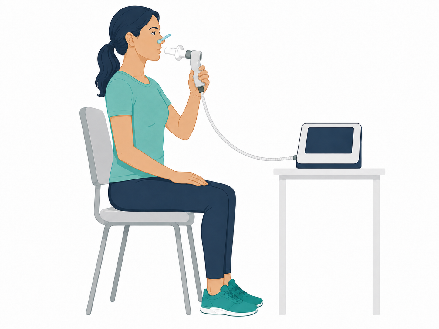
Spirometry does two jobs well: it tells us whether the pattern of your breathing is normal, obstructed or restricted, and it gives a baseline that can be tracked over time and compared before and after treatment.
What spirometry measures
From a single forced breath out, the spirometer records a handful of key values. The three that matter most are FVC, FEV1 and the ratio between them.
| Measure | What it is | What it tells us |
|---|---|---|
| FVC Forced Vital Capacity | The total volume of air you can forcibly breathe out after taking the deepest breath in possible. | An overall measure of lung size and emptying. A low FVC suggests the lungs are holding less air than expected (a restrictive picture), or that air is trapped and cannot be fully exhaled. |
| FEV1 Forced Expiratory Volume in 1 second | The volume of air you blow out in the first second of that forced breath. | A measure of how freely air flows out of the lungs. FEV1 falls when the airways are narrowed, and it is used to grade the severity of airflow obstruction and to monitor change over time. |
| FEV1/FVC ratio | FEV1 expressed as a proportion of FVC — the fraction of your total exhaled air that comes out in the first second. | The single most useful number for distinguishing the type of breathing problem. A reduced ratio points to obstruction; a preserved or raised ratio points to restriction. |
Results are compared against predicted values for someone of the same age, height, sex and ethnic background. The current Australian standard for primary care — the TSANZ Technical Standards for Spirometry in Australian Primary Care (SPC01, 2026) — requires Global Lung Function Initiative (GLI) 2012 reference values and interpretation by z-score rather than by percentage of predicted.1 A z-score simply states how far a result sits from the average for that reference population, in standard deviations: the lower limit of normal (LLN) is a z-score of −1.65, the 5th percentile of healthy people.
This matters clinically. The familiar fixed cut-off of an FEV1/FVC ratio below 0.70 over-diagnoses obstruction in older people, whose ratio falls naturally with age, and under-diagnoses it in the young.1,2 A z-score adjusts for age automatically, so the same threshold carries the same meaning at 25 and at 75.
How results are graded
Under SPC01, airflow obstruction is present when the FEV1/FVC ratio is below the LLN (z-score below −1.65). Where impairment is graded, it is graded on the z-score, in three tiers:1
| Impairment | Z-score range | What it means in practice |
|---|---|---|
| Mild | −1.65 to −2.5 | Below the normal range, but only modestly. Often asymptomatic at rest. |
| Moderate | −2.51 to −4 | Clearly reduced. Usually associated with breathlessness on exertion. |
| Severe | Below −4.1 | Markedly reduced, with substantial limitation of daily activity in most people. |
Two cautions travel with this table, and SPC01 states both explicitly. Lung function sits on a continuum, so severity labels should be applied with care near their boundaries. And lung function severity is not disease severity: symptoms, functional capacity, imaging and comorbidities all belong in the overall assessment.1
The older approach of grading by percentage of predicted FEV1 (70%, 60%, 50%, 35%) is no longer recommended for impairment grading, because it does not grade consistently across ages and misclassifies older adults in particular.1 Percentage of predicted still appears in disease-specific staging systems — GOLD grades 1–4 in COPD, for instance — and those remain in use for that purpose.3
Obstructive vs restrictive patterns
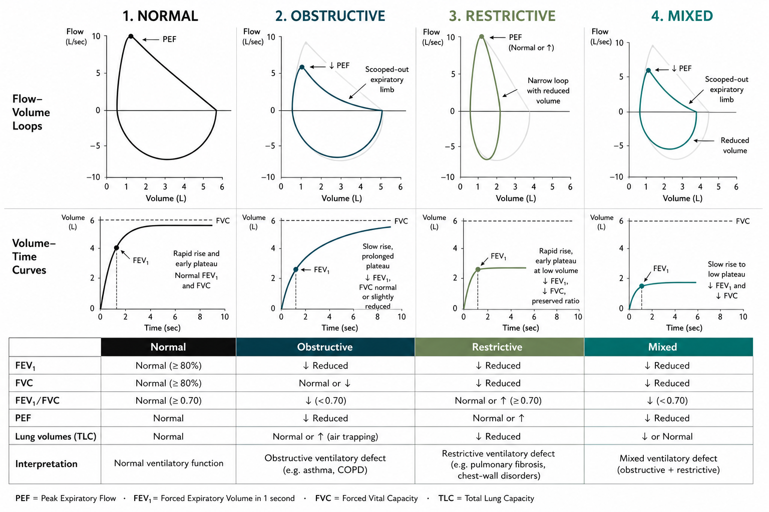
Putting the three values together gives the overall pattern, which guides what the problem is likely to be. The four classic patterns — normal, obstructive, restrictive and mixed — are shown side by side in Figure 2.
Normal. FEV1, FVC and the FEV1/FVC ratio are all within the expected range (all z-scores above −1.65). Exercise capacity is normal and any breathlessness on exertion is in proportion to fitness — the first column of Figure 2.
Obstructive — asthma, COPD or bronchiectasis. The airways are narrowed, so air leaves the lungs slowly: FEV1 falls more than FVC, pulling the FEV1/FVC ratio below the LLN, which is the hallmark of obstruction (FVC is normal, or reduced where air becomes trapped). FEV1 itself may or may not be below the LLN, depending on how severe the obstruction is.1,4 This produces the “scooped-out” expiratory limb seen in Figure 2. On exertion, airflow limitation and dynamic hyperinflation (air trapping) make capacity ventilation-limited; a pre-exercise bronchodilator, paced or interval-based training and pulmonary rehabilitation improve tolerance.
Restrictive — pulmonary fibrosis, chest-wall or neuromuscular disease, or obesity. The lungs cannot fully expand, so FEV1 and FVC fall together while the FEV1/FVC ratio is preserved or even raised — the tall, narrow loop in Figure 2. Low lung volumes force rapid, shallow breathing and can cause early exertional oxygen desaturation, limiting endurance, so we monitor SpO2, build activity up gradually, and prioritise pacing and breathing efficiency. A restrictive pattern usually needs confirmation with fuller lung function testing (lung volumes), because spirometry measures only what you can blow out — not the total volume the lungs can hold.
A fourth, mixed pattern shows reduced FEV1, FVC and ratio together, combining features of both — the final column of Figure 2.
Bronchodilator responsiveness
If obstruction is found, the test is repeated about 15 minutes after an inhaled reliever, usually four puffs of salbutamol through a spacer. A meaningful improvement (“bronchodilator responsiveness”, BDR) supports asthma; obstruction that changes little is more typical of COPD.5,6 This before-and-after comparison is one of the most useful measurements spirometry can give.
How “meaningful” is defined has changed, and both definitions are still in circulation, so it is worth knowing which one a report is using.
| Definition | Threshold | Status |
|---|---|---|
| Change as % of predicted Thoracic Society of Australia and New Zealand (TSANZ) SPC01, 2026 | Increase of >10% of the predicted value in FEV1 or FVC | The current Australian standard for primary care. Because the denominator is the predicted value rather than the patient's own baseline, it is not distorted by baseline lung function, height, sex or age.1 |
| Change from baseline the older rule | Increase of ≥12% and ≥200 mL in FEV1 or FVC | Legacy. Still widely quoted and still built into many spirometer printouts, but methods based on absolute change or percentage change from baseline are no longer recommended for defining BDR.1 |
The two are not interchangeable, and SPC01 makes the point directly: a change of more than 10% of predicted is not the same as a 10% change from baseline.1 A patient with small lungs can clear the old percentage threshold on a tiny absolute gain, while a patient with near-normal predicted values may improve substantially and still not reach it.
A reported BDR should therefore state the pre- and post-bronchodilator FEV1 and FVC, and express the change as a percentage of predicted. Responsiveness in FVC rather than FEV1 may reflect a reduction in air trapping, and is interpreted in that light.1
One caveat that applies to both definitions: BDR is a single measurement on a single day. A negative test does not exclude asthma, and a positive test does not confirm it — clinical context always governs.
How the test is done
You will be asked to sit upright, often wearing a soft nose clip, and to seal your lips around a mouthpiece with your tongue underneath it. After breathing normally, you take the biggest breath in you can, then blast the air out as hard and as long as you can — you will be coached to keep going until the flow has all but stopped, rather than to a fixed number of seconds — and then breathe in fully again.
The manoeuvre is repeated until at least three acceptable blows have been recorded, and again after a bronchodilator if responsiveness is being tested.1 Coaching between attempts is not a sign that something is wrong; it is how a good test is produced. Most technical problems — a slow start, a cough in the first second, a leak at the lips, the tongue blocking the mouthpiece, stopping early — are fixed by re-instruction and another attempt.1
You may be asked to avoid bronchodilator inhalers, heavy meals, vigorous exercise, caffeine, smoking and alcohol for a period beforehand, as these can affect the result — your testing service will give you specific instructions.
What makes a test good enough to act on
Spirometry is effort-dependent, so the numbers are only as good as the blows that produced them. SPC01 sets explicit criteria, adopted from the ATS/ERS technical standard, and a report should say whether they were met.1,7
An acceptable blow
Every one of the following must hold:
- A full breath in before the forced blow out.
- A rapid start — back-extrapolated volume (BEV) no more than 5% of FVC or 0.10 L, whichever is greater.
- No cough, leak or artefact during the manoeuvre.
- A continuous maximal blow reaching one end-of-test criterion: a plateau on the volume–time curve (a change of 0.025 L or less over the final second), or an expiratory time of 15 seconds or more, or an FVC within 0.15 L of the largest previous FVC where no plateau is achievable.
- Where an inspiratory loop is recorded, FIVC must not exceed FVC by more than 0.10 L or 5%.
A repeatable session
The session is repeatable when the two largest acceptable FEV1 values are within 0.15 L of each other, and the two largest acceptable FVC values likewise.1
Quality grade
Acceptability and repeatability combine into a letter grade, assigned separately for FEV1 and FVC, and separately before and after a bronchodilator. Grade A or B is the minimum standard for primary-care testing: Grade A means three or more acceptable blows meeting repeatability, Grade B means two.1 Lower grades are not automatically discarded — they can still carry clinically useful information — but the grade has to be documented and the interpretation qualified accordingly.
Alongside the grade, the operator records the medication taken before the test, the bronchodilator given and the interval before retesting, observations on effort and technique, and whether acceptability and repeatability were met. The report shows the largest FEV1 and largest FVC, with the flow–volume and volume–time curves from three acceptable manoeuvres.1
Tracking change over time
Where earlier tests exist, results are compared with them — and a sustained fall matters as much as a single low reading. A decline in FEV1 or FVC of more than 15% from baseline in GLI percentage of predicted exceeds expected year-to-year variability and warrants further evaluation, whether from disease progression, treatment failure, or workplace exposure.1
For this comparison to mean anything, the same reference category must be used on every occasion — which is why a change of assigned ethnicity has to be flagged on the report.
Who should perform it
SPC01 is a technical standard for the test; a companion standard, SPC02, sets the training and competency requirements for the people performing it, including initial training and ongoing maintenance of competency.8 In Queensland, spirometry performed for statutory mine dust lung disease surveillance must be done by providers approved against both standards.
Why it matters in cardiorespiratory physiotherapy
Spirometry helps confirm a diagnosis, grade its severity, and measure whether treatment is working. For physiotherapy, the pattern and severity of any obstruction or restriction inform which techniques are likely to help, how hard to push during exercise, and how to track progress over time. It also flags when onward referral for fuller lung function testing or specialist review is needed.
References & evidence base
- Thoracic Society of Australia and New Zealand. SPC01: TSANZ Technical Standards for Spirometry in Australian Primary Care. TSANZ; 2026.
- Stanojevic S, Kaminsky DA, Miller MR, et al. ERS/ATS technical standard on interpretive strategies for routine lung function tests. Eur Respir J 2022;60(1):2101499.
- Global Initiative for Chronic Obstructive Lung Disease. Global strategy for the diagnosis, management and prevention of COPD: 2026 report. GOLD; 2026. Available at: goldcopd.org
- Quanjer PH, Stanojevic S, Cole TJ, et al. Multi-ethnic reference values for spirometry for the 3–95-yr age range: the global lung function 2012 equations. Eur Respir J 2012;40(6):1324–1343.
- National Asthma Council Australia. Australian Asthma Handbook. Version 3.0. Melbourne: National Asthma Council Australia; 2025. Available at: asthmahandbook.org.au
- National Institute for Health and Care Excellence. Asthma: diagnosis, monitoring and chronic asthma management (NG80). London: NICE, 2019.
- Graham BL, Steenbruggen I, Miller MR, et al. Standardization of spirometry 2019 update: an official American Thoracic Society and European Respiratory Society technical statement. Am J Respir Crit Care Med 2019;200(8):e70–e88.
- Thoracic Society of Australia and New Zealand. SPC02: TSANZ Standards for Training and Maintaining Competency in Spirometry for Australian Primary Care. TSANZ; 2026.
References are numbered in citation order (Vancouver/BMJ style) and were current at the time of writing. Guidelines are living documents — verify against the latest version before clinical use.
We perform spirometry as part of your assessment and use the results to plan and review your treatment.
Corrections: If something on this page is wrong, out of date or unclear, we want to know. Email reception@inspireclinic.au with the page name and what you believe is incorrect. Substantive corrections are made promptly, and the guide’s version and last-updated date are changed to reflect it.