Ahpra registration PHY0002298174
How these guides are written and reviewed →
In short
- Call 000 now: Coughing up more than a teaspoon of blood — or any blood together with breathlessness, light-headedness or feeling faint.
- Go to the emergency department today: Breathlessness at rest that is new or much worse than your usual level, chest pain on breathing in, or fever with shivering, confusion or drowsiness.
- See a doctor the same day: An exacerbation: more sputum than usual or a change in its colour, worsening cough or breathlessness lasting more than about 48 hours, or unusual fatigue and feeling unwell.
- A physiotherapist teaches you an airway clearance routine that suits you, fits it around your saline nebuliser, and checks your technique regularly.
Bronchiectasis is a condition where the airways become permanently widened and scarred, so mucus pools and is hard to clear. This leads to a chronic cough with phlegm and repeated chest infections, which can cause further damage — a cycle we aim to break. Day-to-day management centres on clearing the airways every day, treating infections promptly, staying active and keeping vaccinations up to date. Nebulised salty water (hypertonic saline) and airway clearance techniques are mainstays. This page explains bronchiectasis and how it is managed.
Bronchiectasis is a chronic suppurative airway disease characterised by abnormal, permanent dilation of the bronchi, with chronic cough, sputum production, and recurrent infective exacerbations. It is increasingly recognised in Australia and forms a core area of cardiorespiratory physiotherapy practice.
Definition
Bronchiectasis is a chronic lung condition characterised by abnormal and permanent dilation of the bronchi, with destruction of the bronchial wall. It is defined radiologically on high-resolution computed tomography (HRCT) by bronchial dilation in relation to the accompanying pulmonary artery, lack of normal airway tapering, and visibility of airways within 1 cm of the pleura.
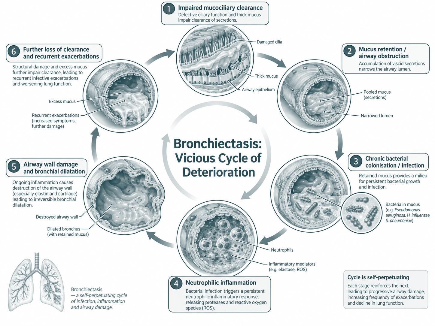
It is the end result of a vicious cycle in which infection, inflammation, impaired mucociliary clearance and structural airway damage perpetuate one another.1
Pathophysiology
The Cole vicious cycle hypothesis describes how an initial insult impairs mucociliary clearance, allowing chronic bacterial colonisation. Persistent bacterial presence triggers a neutrophil-dominated inflammatory response, releasing proteases (notably neutrophil elastase) that damage the airway wall. This worsens clearance, perpetuating colonisation, inflammation, and progressive structural damage.
Co-morbidities
Common co-morbidities include chronic rhinosinusitis, gastro-oesophageal reflux, cardiovascular disease, anxiety and depression, urinary incontinence (from chronic cough), and osteoporosis. Co-existing chronic obstructive pulmonary disease (COPD) and asthma are also frequent.
Prevalence
Bronchiectasis is increasingly recognised in Australia. Prevalence rises sharply with age, and it is over-represented in Aboriginal and Torres Strait Islander people, in whom childhood-onset bronchiectasis remains a significant health burden. International prevalence estimates range from 67 to over 1,100 per 100,000, with the higher figures from older populations.
Causes
Causes are heterogeneous and a structured aetiological work-up identifies a cause in 50–75% of adults. Common identifiable causes include:
- Post-infectious (childhood pneumonia, tuberculosis, whooping cough, measles).
- Immunodeficiency (common variable immunodeficiency, IgG subclass deficiency, HIV).
- Cystic fibrosis and CFTR-related disease.
- Primary ciliary dyskinesia.
- Alpha-1 antitrypsin deficiency.
- Allergic bronchopulmonary aspergillosis (ABPA).
- Rheumatological disease (rheumatoid arthritis, Sjögren’s, IBD-related).
- Aspiration and gastro-oesophageal reflux.
- Non-tuberculous mycobacterial infection.
- Idiopathic (no cause identified after work-up).
Symptoms
The hallmark symptoms are chronic productive cough, daily sputum (often purulent), recurrent chest infections, breathlessness, fatigue, chest pain, and haemoptysis. Many patients experience progressive exercise limitation and reduced quality of life. Symptom burden does not always correlate with radiological severity.
Nontuberculous mycobacteria (NTM)
NTM, particularly Mycobacterium avium complex (MAC) and M. abscessus, are increasingly recognised in patients with bronchiectasis. NTM should be considered in patients with worsening symptoms despite optimised therapy, declining lung function, new nodular or tree-in-bud changes on HRCT, or persistent positive sputum cultures. Diagnosis and management require specialist respiratory input and meet ATS/ERS/ESCMID/IDSA criteria.
Diagnosis
Importance of a diagnosis
A formal diagnosis enables targeted treatment, identification of a treatable underlying cause, access to appropriate antibiotic and airway clearance regimens, and access to peer support and resources. Untreated bronchiectasis leads to progressive lung damage, recurrent infections, and reduced quality of life.
How is it diagnosed?
Diagnosis is made on HRCT in a patient with appropriate clinical symptoms (chronic productive cough, recurrent chest infections). The diagnostic process should include a systematic search for an underlying cause, sputum microbiology, lung function testing, and assessment of disease severity.2,3
Radiology
HRCT is the gold-standard imaging modality. Features include bronchial dilation greater than the accompanying pulmonary artery (signet-ring sign), lack of bronchial tapering, visibility of airways within 1 cm of the pleura, and bronchial wall thickening. Mucous plugging, tree-in-bud nodules, and mosaic attenuation are common associated findings. Chest X-ray is insensitive and should not be relied upon to exclude bronchiectasis.
Lung function
Spirometry typically shows an obstructive pattern, though restrictive and mixed patterns occur. FEV1 is used to track disease progression. Full lung function (lung volumes and DLCO) is informative when there is co-existing emphysema or fibrosis. Lung function does not correlate strongly with radiological severity.
Sputum pathology
Baseline and periodic sputum culture (with sensitivities) is recommended, including specific testing for Pseudomonas aeruginosa, NTM, and fungi. Common organisms include Haemophilus influenzae, P. aeruginosa, S. aureus, Moraxella catarrhalis, and S. pneumoniae. Chronic Pseudomonas infection is associated with worse outcomes and warrants attempted eradication.
Investigations for secondary causes
A standard work-up may include: serum immunoglobulins (IgG, IgA, IgM and IgG subclasses), specific antibody responses to vaccines, HIV status (when appropriate), alpha-1 antitrypsin, sweat chloride and/or CFTR genetic testing (in selected patients), aspergillus precipitins and specific IgE for ABPA, rheumatological serology when clinical features are present, and ciliary studies in patients with suggestive features.
Management
Management and goals
The goals of bronchiectasis management are to: reduce exacerbations, improve symptoms (cough, sputum, breathlessness), preserve lung function, improve exercise capacity and quality of life, and address underlying contributors.
Treatment options
Treatment is multimodal and tailored to the individual. Core components include:
- Airway clearance therapy (the cornerstone of management).
- Exercise and pulmonary rehabilitation.
- Treatment of acute exacerbations with appropriate antibiotics.
- Long-term macrolide therapy (e.g. azithromycin) for frequent exacerbators — sputum must be cultured for nontuberculous mycobacteria before it starts, because macrolide monotherapy in unrecognised NTM infection induces resistance and compromises later treatment.
- Eradication of newly isolated Pseudomonas aeruginosa.
- Inhaled antibiotics in selected chronic Pseudomonas infection.
- Mucoactive therapy (hypertonic saline, e.g. PARI MucoClear® 3–6%; dornase alfa is generally avoided as it has been shown to worsen outcomes in non-CF bronchiectasis).
- Vaccination (influenza, pneumococcal, COVID-19, pertussis).
- Management of the underlying cause and co-morbidities.4,5,6,7,8,9
Identifying an exacerbation
An exacerbation is defined by deterioration of three or more key symptoms for ≥48 hours: cough, sputum volume or consistency, sputum purulence, breathlessness, exercise tolerance, fatigue or malaise, or haemoptysis. The clinician should also note any systemic features (fever, weight loss) or oxygen desaturation.
Action plan
A written, individualised action plan helps the patient recognise the early signs of an exacerbation and initiate treatment promptly. It typically includes baseline symptoms (the “well zone”), instructions for increasing airway clearance, when to start antibiotics (usually a 14-day course based on prior sputum sensitivities), and clear criteria for contacting the GP, physiotherapist, respiratory specialist, or attending hospital.
Medications
Medications for bronchiectasis
Common medication classes include short- and long-acting bronchodilators (where airflow obstruction or reversibility is present), inhaled corticosteroids (only when co-existing asthma or eosinophilic inflammation is documented), nebulised hypertonic saline, long-term macrolides (azithromycin), oral and inhaled antibiotics for exacerbations and chronic infection, and vaccinations.
Correct use of medications
Inhaler and nebuliser technique should be reviewed at every encounter, as poor technique substantially reduces drug delivery and efficacy. Patients should be taught the difference between rescue and maintenance medications, and the rationale for adherence even when feeling well.
Order of medications
When multiple inhaled or nebulised therapies are used, the recommended order is:
- Bronchodilator (to open the airways)
- Mucoactive therapy (hypertonic saline) to mobilise secretions
- Airway clearance technique
- Inhaled antibiotic (if prescribed) to maximise deposition on cleared airways
- Inhaled corticosteroid (if indicated)
Extra-pulmonary manifestations
Sinusitis
Chronic rhinosinusitis is present in up to 70% of patients with bronchiectasis and may share common underlying mechanisms (e.g. primary ciliary dyskinesia, immunodeficiency). Saline nasal irrigation, intranasal corticosteroids, and ENT review where indicated form the mainstay of management.
Gastro-oesophageal reflux
GORD is both a potential cause and a perpetuator of bronchiectasis through micro-aspiration. Lifestyle measures (weight management, elevation of the head of the bed, meal timing) and proton pump inhibitors may be helpful; specialist review is warranted in refractory cases.
Urinary incontinence
Stress urinary incontinence is highly prevalent in adults with bronchiectasis (particularly women) due to chronic cough. Pelvic floor physiotherapy and bladder training are first-line interventions and should be offered routinely.
Musculoskeletal issues
Chronic cough and altered breathing mechanics commonly produce thoracic spine stiffness, intercostal and accessory muscle dysfunction, rib pain, and posture-related shoulder pain. Manual therapy, mobility work, postural retraining, and breathing pattern correction are useful adjuncts.
Living with bronchiectasis
Nutrition
Adequate energy and protein intake supports immune function and muscle mass. Weight loss and sarcopenia are associated with worse outcomes. Hydration is important for mucus clearance. Dietitian referral is recommended where intake is suboptimal or there is unintended weight loss.
Sleep
Cough, sputum and breathlessness disrupt sleep. Patients should be screened for obstructive sleep apnoea where appropriate. A pre-bed airway clearance routine may improve sleep quality.
Travel
Most people with bronchiectasis can travel safely. Considerations include vaccinations, carrying a sufficient supply of medications and a recent prescription, a clinician letter detailing the diagnosis and equipment (nebulisers, oscillating positive expiratory pressure (PEP) devices), travel insurance that covers the condition, and assessment for in-flight oxygen if resting SpO₂ is below 92–95% or if there is significant gas exchange impairment.
Prognosis
Prognosis varies with the underlying cause, exacerbation frequency, chronic infection (particularly P. aeruginosa), lung function, and co-morbidities. Validated severity scores include the Bronchiectasis Severity Index (BSI) and the FACED score, both of which predict mortality and hospitalisation risk.10,11
Anxiety and depression
Anxiety and depression are common in chronic lung disease and are under-recognised. Screening (e.g. HADS, PHQ-9, GAD-7) should be incorporated into routine review, with referral for psychological support, peer support groups, and pharmacological treatment when indicated.
Role of the physiotherapist
Physiotherapy is the cornerstone of day-to-day bronchiectasis care. The cardiorespiratory physiotherapist selects and teaches an individualised airway clearance routine (for example ACBT or an oscillating device), times it with nebulised hypertonic saline, and reviews technique and adherence regularly. They also lead exercise and pulmonary rehabilitation, educate on recognising and acting on a flare-up, and address related problems such as cough-related incontinence and breathing pattern.12,13,14
Warning signs
Part 1 · References
- Flume PA, Chalmers JD, Olivier KN. Advances in bronchiectasis: endotyping, genetics, microbiome, and disease heterogeneity. Lancet 2018;392(10150):880–890.
- Polverino E, Goeminne PC, McDonnell MJ, et al. European Respiratory Society guidelines for the management of adult bronchiectasis. Eur Respir J 2017;50(3):1700629.
- Hill AT, Sullivan AL, Chalmers JD, et al. British Thoracic Society guideline for bronchiectasis in adults. Thorax 2019;74(Suppl 1):1–69.
- Nicolson CH, Stirling RG, Borg BM, Starkey CR, Thompson PJ, Holmes PW. The long term effect of inhaled hypertonic saline 6% in non-cystic fibrosis bronchiectasis. Respir Med 2012;106(5):661–667.
- Bilton D, Tino G, Barker AF, et al. Inhaled mannitol for non-cystic fibrosis bronchiectasis: a randomised, controlled trial. Thorax 2014;69(12):1073–1079.
- O'Donnell AE, Barker AF, Ilowite JS, Fick RB. Treatment of idiopathic bronchiectasis with aerosolized recombinant human DNase I. Chest 1998;113(5):1329–1334.
- Serisier DJ, Martin ML, McGuckin MA, et al. Effect of long-term, low-dose erythromycin on pulmonary exacerbations among patients with non-cystic fibrosis bronchiectasis: the BLESS randomized controlled trial. JAMA 2013;309(12):1260–1267.
- Wong C, Jayaram L, Karalus N, et al. Azithromycin for prevention of exacerbations in non-cystic fibrosis bronchiectasis (EMBRACE): a randomised, double-blind, placebo-controlled trial. Lancet 2012;380(9842):660–667.
- Chalmers JD, Burgel PR, Daley CL, et al; ASPEN Investigators. Phase 3 trial of the DPP-1 inhibitor brensocatib in bronchiectasis. N Engl J Med 2025;392(16):1569–1581. Supersedes the 2020 phase 2 trial.
- Chalmers JD, Goeminne P, Aliberti S, et al. The bronchiectasis severity index: an international derivation and validation study. Am J Respir Crit Care Med 2014;189(5):576–585.
- Finch S, McDonnell MJ, Abo-Leyah H, Aliberti S, Chalmers JD. A comprehensive analysis of the impact of Pseudomonas aeruginosa colonization on prognosis in adult bronchiectasis. Ann Am Thorac Soc 2015;12(11):1602–1611.
- Lee AL, Burge AT, Holland AE. Airway clearance techniques for bronchiectasis. Cochrane Database Syst Rev 2015;(11):CD008351.
- Muñoz G, de Gracia J, Buxó M, Alvarez A, Vendrell M. Long-term benefits of airway clearance in bronchiectasis: a randomised placebo-controlled trial. Eur Respir J 2018;51(1):1701926.
- Lee AL, Hill CJ, Cecins N, et al. The short and long term effects of exercise training in people with non-cystic fibrosis bronchiectasis: a randomised controlled trial. Respir Res 2014;15:44.
References are numbered in citation order (Vancouver/BMJ style) and were current at the time of writing. Guidelines are living documents — verify against the latest version before clinical use.
Clearing mucus well is a skill rather than a machine. We match a technique to your lungs and your routine, then coach it until you can do it at home on a bad morning.
Clinical evidence
Part 1 covers the same condition without the technical detail. What follows is the evidence base behind it, written for clinicians — the literature, the reasoning and the gaps.
For clinicians: this summary supports clinical reasoning and is not a protocol. Check current guidelines and local policy before applying it, and read it alongside the key references and guidelines directory.
Framing. Bronchiectasis is understood as a self-perpetuating "vicious vortex" of impaired mucociliary clearance, chronic infection, inflammation and structural damage, each element driving the others.1 Airway clearance sits deliberately at the centre of both the ERS and BTS guidelines — not because the randomised evidence is large, but because it is the only intervention that acts directly on the mucus limb of that cycle, and because the aetiological and microbiological workup around it changes management in a substantial minority of patients.2,3
Airway clearance and mucoactive therapy
- Airway clearance techniques improve sputum expectoration and health-related quality of life across small trials, with no technique demonstrating consistent superiority; the Cochrane review found no adequately powered evidence for an effect on exacerbation frequency.4
- ELTGOL performed twice daily for a year reduced exacerbations and improved quality of life versus sham in a randomised placebo-controlled trial — the strongest single trial supporting sustained daily clearance.5
- Hypertonic saline 6% improved quality of life and lung function over 12 months, but not more than isotonic saline when both were added to a supervised clearance regimen — the technique, not the tonicity, may carry much of the effect.6 Inhaled mannitol improved quality of life but did not significantly reduce exacerbation rate.7
- rhDNase (dornase alfa) is harmful in bronchiectasis — more exacerbations and greater FEV1 decline than placebo. It is a cystic fibrosis drug and must not be extrapolated.8
Exercise, antibiotics and prognosis
- Pulmonary rehabilitation improves exercise capacity and dyspnoea and reduced exacerbation frequency over 12 months, although the exercise-capacity gain was not maintained at that point without ongoing training — an argument for maintenance rather than a single course.9
- Long-term macrolides (BLESS, EMBRACE) reduce exacerbation frequency in patients with recurrent exacerbations, after excluding non-tuberculous mycobacteria; inhaled antibiotics are used in chronic Pseudomonas aeruginosa infection.10,11
- Severity and prognosis are best stratified with the Bronchiectasis Severity Index; chronic P. aeruginosa infection independently predicts exacerbations, hospitalisation and mortality.12,13
- Anti-inflammatory therapy targeting neutrophil serine proteases (brensocatib, DPP-1 inhibition) reduced exacerbations in the phase 3 ASPEN trial — 1,721 patients, roughly a 20% reduction in the annualised exacerbation rate at both doses, with a slower rate of FEV1 decline at 25 mg — opening a treatment axis that does not depend on antibiotics. It became the first approved therapy for bronchiectasis in the United States in August 2025.14 It is not currently approved by the TGA and is not PBS-listed in Australia for non-cystic-fibrosis bronchiectasis, so it is not an available treatment option here — worth knowing, because patients increasingly arrive having read about it.
Physiotherapy implications
- Adherence outweighs technique selection. With no clear winner among active cycle of breathing techniques (ACBT), ELTGOL, autogenic drainage and oscillating PEP, choose on sputum volume, dexterity, fatigue, home setting and patient preference, then review the technique in person as disease and adherence change.
- Sequence nebulised therapy correctly: bronchodilator → hypertonic saline → airway clearance → nebulised antibiotic last, so the antibiotic deposits on cleared airways. Cover the first hypertonic saline dose with a reliever and a spirometric or symptomatic check for bronchoconstriction.
- Treat exercise as exacerbation-modifying therapy, not general fitness advice, and plan a maintenance pathway from the outset given the 12-month attenuation.9
- Screen for cough-related urinary incontinence — common in women with chronic cough and a frequent, unvolunteered reason for avoiding huffing and coughing at home.3
- Modify during frank haemoptysis: defer percussion, vigorous huffing and head-down positioning until the bleeding is assessed and settled; resume graded clearance rather than abandoning it.
- Infection control matters: single-patient devices, taught cleaning and drying, and segregation practices where P. aeruginosa or NTM are present.
Clinical reasoning
- A patient who is "not clearing" usually has a dose, timing or hydration problem rather than needing a new device — audit the routine before changing it.
- Rising sputum volume, purulence change or new haemoptysis is an exacerbation trigger for medical review; escalate rather than intensifying clearance alone.
- Persistent failure to respond, or an atypical microbiology profile, should prompt reconsideration of NTM — which changes both antibiotic strategy and macrolide eligibility.
- Suspected undiagnosed cystic fibrosis, primary ciliary dyskinesia, immunodeficiency or ABPA in a younger or refractory patient warrants aetiological workup, because it changes treatment.2
Evidence gaps
- No adequately powered trial compares airway clearance techniques head-to-head against exacerbation outcomes; dose and frequency remain guideline consensus rather than evidence.
- Optimal maintenance strategy after pulmonary rehabilitation is unestablished.
- Whether mucoactive agents add benefit beyond a well-taught clearance routine remains unresolved.6
- Most trial cohorts are stable, ambulatory adults; the frail, the elderly and those with severe airflow obstruction are under-represented.
References for the clinical evidence summary
- Flume PA, Chalmers JD, Olivier KN. Advances in bronchiectasis: endotyping, genetics, microbiome, and disease heterogeneity. Lancet 2018;392(10150):880–890.
- Polverino E, Goeminne PC, McDonnell MJ, et al. European Respiratory Society guidelines for the management of adult bronchiectasis. Eur Respir J 2017;50(3):1700629.
- Hill AT, Sullivan AL, Chalmers JD, et al. British Thoracic Society guideline for bronchiectasis in adults. Thorax 2019;74(Suppl 1):1–69.
- Lee AL, Burge AT, Holland AE. Airway clearance techniques for bronchiectasis. Cochrane Database Syst Rev 2015;(11):CD008351.
- Muñoz G, de Gracia J, Buxó M, Alvarez A, Vendrell M. Long-term benefits of airway clearance in bronchiectasis: a randomised placebo-controlled trial. Eur Respir J 2018;51(1):1701926.
- Nicolson CH, Stirling RG, Borg BM, Starkey CR, Thompson PJ, Holmes PW. The long term effect of inhaled hypertonic saline 6% in non-cystic fibrosis bronchiectasis. Respir Med 2012;106(5):661–667.
- Bilton D, Tino G, Barker AF, et al. Inhaled mannitol for non-cystic fibrosis bronchiectasis: a randomised, controlled trial. Thorax 2014;69(12):1073–1079.
- O'Donnell AE, Barker AF, Ilowite JS, Fick RB. Treatment of idiopathic bronchiectasis with aerosolized recombinant human DNase I. Chest 1998;113(5):1329–1334.
- Lee AL, Hill CJ, Cecins N, et al. The short and long term effects of exercise training in people with non-cystic fibrosis bronchiectasis: a randomised controlled trial. Respir Res 2014;15:44.
- Serisier DJ, Martin ML, McGuckin MA, et al. Effect of long-term, low-dose erythromycin on pulmonary exacerbations among patients with non-cystic fibrosis bronchiectasis: the BLESS randomized controlled trial. JAMA 2013;309(12):1260–1267.
- Wong C, Jayaram L, Karalus N, et al. Azithromycin for prevention of exacerbations in non-cystic fibrosis bronchiectasis (EMBRACE): a randomised, double-blind, placebo-controlled trial. Lancet 2012;380(9842):660–667.
- Chalmers JD, Goeminne P, Aliberti S, et al. The bronchiectasis severity index: an international derivation and validation study. Am J Respir Crit Care Med 2014;189(5):576–585.
- Finch S, McDonnell MJ, Abo-Leyah H, Aliberti S, Chalmers JD. A comprehensive analysis of the impact of Pseudomonas aeruginosa colonization on prognosis in adult bronchiectasis. Ann Am Thorac Soc 2015;12(11):1602–1611.
- Chalmers JD, Burgel PR, Daley CL, et al; ASPEN Investigators. Phase 3 trial of the DPP-1 inhibitor brensocatib in bronchiectasis. N Engl J Med 2025;392(16):1569–1581. Supersedes the 2020 phase 2 trial.
Corrections: If something on this page is wrong, out of date or unclear, we want to know. Email reception@inspireclinic.au with the page name and what you believe is incorrect. Substantive corrections are made promptly, and the guide’s version and last-updated date are changed to reflect it.