Ahpra registration PHY0002298174
How these guides are written and reviewed →
Endobronchial valves for severe, hyperinflated emphysema: selection criteria (target-lobe destruction, intact fissures or absent collateral ventilation, hyperinflation), the procedure, pneumothorax risk after insertion, trial outcomes, and physiotherapy before and after. Client information follows in the boxed sections.
In severe emphysema the worst-damaged part of the lung fills with trapped air and presses on the healthier lung underneath. Zephyr valves are tiny one-way valves placed through a camera down the airway — no cuts, no chest opening. They let air out of the target lobe but not back in, so that lobe slowly deflates and the better lung, the diaphragm and the ribcage get room to work again. Chosen well, people breathe more easily, walk further and feel better; chosen badly, the valves do nothing and carry real risk. The selection is everything.
This guide explains what bronchoscopic lung volume reduction (BLVR) with endobronchial valves is, who it suits, what the procedure and recovery involve, and where physiotherapy fits before and after. A clinician evidence summary follows the patient section — jump to it.
Why trapped air is the problem
Emphysema destroys the elastic walls between air sacs. The lung loses its spring, so air goes in but struggles to come out. Over time the worst-affected lobes become permanently over-inflated — hyperinflation — which:
- pushes the diaphragm flat, taking away its most powerful range of movement;
- squeezes the healthier lung tissue that is still capable of gas exchange;
- forces you to start each breath with your lungs already part-full, so the work of breathing climbs steeply with activity;
- makes breathlessness worse than the airflow numbers alone would predict.
Bronchodilators, pulmonary rehabilitation and breathing retraining all help you live better with hyperinflation. Volume reduction is different: it physically removes the over-inflated lobe from the equation.
What Zephyr valves are, and what they do
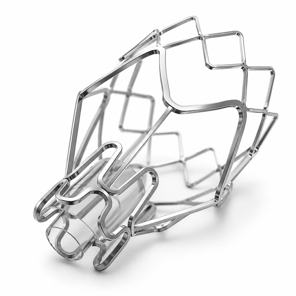
A Zephyr valve is a small silicone-and-nitinol one-way valve, about the size of a grain of rice, placed inside an airway. Several (typically three to five) are placed in the airways feeding one target lobe. The valve closes on the way in and opens on the way out, so:
Who is suitable
Valves work only in a narrow group, and the assessment is deliberately thorough. A specialist respiratory team decides — usually at a multidisciplinary meeting — using breathing tests, a high-resolution CT and often a measurement taken during bronchoscopy.1,2
- Severe emphysema with marked hyperinflation on lung-function testing (a high residual volume is the key number, not just a low FEV1)
- Breathlessness that still limits daily life despite optimised inhalers, oxygen where indicated, and a completed course of pulmonary rehabilitation
- Emphysema that is uneven — one lobe clearly worse than the rest, so there is a sensible target
- Little or no collateral ventilation — the target lobe must not be fed with air leaking around from its neighbour (assessed by fissure integrity on CT and/or a Chartis measurement at bronchoscopy)
- Stopped smoking, and able to cooperate with the procedure and recovery
- Collateral ventilation is present — the lobe will not deflate, so valves cannot work
- Frequent infective exacerbations, active infection, or significant bronchiectasis with heavy sputum in the target area
- Very poor gas transfer, severe pulmonary hypertension or significant heart disease that makes anaesthetic and recovery unsafe
- Large bullae, previous major lung surgery or pleurodesis in the target region
- Still smoking, or unable to attend follow-up
An important note about expectations. Valves do not cure emphysema and do not stop it progressing. They change the mechanics of breathing. Some people improve markedly, some modestly, and some not at all — which is why the team spends so long on selection before offering the procedure.
What happens — the procedure and the hospital stay
What the results look like
In the pivotal trial of Zephyr valves in carefully selected patients, average results at 12 months compared with continued medical care were:1
Averages hide individuals. Ask your specialist what the realistic range is for your CT and lung volumes.
Where physiotherapy fits
Before the procedure
- Pulmonary rehabilitation is part of the pathway, not an optional extra. Programmes are expected before valves are offered, and fitter patients tolerate the anaesthetic and the recovery better.2
- Airway clearance and sputum control. A wet, poorly cleared chest raises infection risk and can rule the procedure out. We optimise clearance before you go in — see Principles of Airway Clearance.
- Inhaler technique and adherence. Valves are offered when medical therapy is optimised; technique errors quietly undermine that. See Getting Your Inhaler Technique Right.
- Breathing pattern and pacing. Practising pursed-lip breathing, recovery positions and pacing before surgery means you already own the tools when you need them afterwards.
- Smoking cessation and nutrition support — both are prerequisites and both change outcome. See Smoking Cessation.
After the procedure
- Early mobilisation in hospital, guided by the treating team, with close attention to any change in breathlessness or pain.
- Airway clearance is re-planned, not simply resumed. The target lobe is deliberately collapsed, so clearance now targets the remaining lung. Techniques that generate high pressures or vigorous coughing are re-introduced cautiously in the early weeks, in consultation with the treating respiratory team — there is no benefit in stressing a fresh valve or a lung at risk of air leak.
- Positive-pressure devices and NIV need a specialist decision. If you use continuous positive airway pressure (CPAP), bilevel positive airway pressure (BiPAP) or a positive expiratory pressure (PEP) device, ask the team explicitly when and how to restart. Do not resume on assumption.
- Pulmonary rehabilitation again, once cleared. This is where the mechanical gain becomes a functional gain: the valves give you room to breathe, rehabilitation converts it into walking distance, stair tolerance and confidence.
- Ongoing review. Benefit can fade if a valve migrates or a new exacerbation pattern develops; we track walking distance and symptom scores over time and flag changes to your specialist.
- Rehabilitation before and after is what turns a successful procedure into a better life.
- Know the pneumothorax warning signs, and carry a note that you have valves in place.
- Never restart positive-pressure therapy or vigorous clearance after valves without asking the team.
Other volume-reduction options
| Option | How it works | Where it sits |
|---|---|---|
| Endobronchial valves (Zephyr; Spiration/IBV) | One-way valves deflate a target lobe; removable | The best-evidenced bronchoscopic option in emphysema without collateral ventilation1,3,4 |
| Lung volume reduction surgery | Surgical removal of the worst-affected lung tissue | Effective in selected upper-lobe-predominant disease with low exercise capacity; higher early risk, considered by thoracic surgery5 |
| Endobronchial coils | Nitinol coils retract lung tissue mechanically; work despite collateral ventilation | Modest benefit, limited availability6 |
| Thermal vapour ablation / sealants | Deliberate scarring shrinks the target segment | Segmental targeting; specialist centres, more limited evidence7 |
| Lung transplantation | Replacement of diseased lung | For younger patients with very advanced disease; separate pathway, strict criteria |
How to explore it in Australia
BLVR is done at selected centres by interventional respiratory physicians, and access starts with a referral from your respiratory specialist — your GP or physiotherapist cannot refer directly for valves. If you think you may fit the picture above, the practical steps are:
- Ask your GP for a respiratory physician review if you do not already have one, and mention that you want your suitability for volume reduction assessed.
- Take your most recent lung-function results and any CT reports to that appointment.
- Complete pulmonary rehabilitation if you have not — it is both a prerequisite and a fair test of what you can gain without a procedure.
- Ask the specialist directly: is my emphysema uneven enough, is my residual volume high enough, and do I have collateral ventilation? Those three answers decide almost everything.
- Clarify costs and pathway — public hospital versus private — before proceeding, as funding arrangements vary by state and by insurer.
Evidence summary — endobronchial valves in severe emphysema
Mechanism and rationale. BLVR targets static hyperinflation rather than airflow obstruction per se. Target-lobe atelectasis reduces residual volume, improves diaphragm configuration and length–tension relationship, and decompresses adjacent, better-perfused parenchyma. The magnitude of symptomatic benefit tracks the degree of target-lobe volume reduction more closely than the change in FEV1.
Trial evidence- STELVIO (randomised, patients selected by Chartis-confirmed absence of collateral ventilation) reported between-group improvements in FEV1, FVC and 6-minute walk distance at 6 months that are among the largest seen with any intervention in severe chronic obstructive pulmonary disease (COPD).3
- TRANSFORM (heterogeneous emphysema, no collateral ventilation) reported a markedly higher proportion of patients achieving a ≥12% FEV1 improvement at 3 months versus standard care, with parallel gains in SGRQ and six-minute walk distance (6MWD).4
- LIBERATE, the 12-month pivotal trial, confirmed durability to one year: mean FEV1 improvement of about 18% over control, +39 m 6MWD, −7.1 points SGRQ, with target-lobe volume reduction the key mediator. Pneumothorax occurred in roughly a quarter of patients, concentrated in the first days.1
- EMPROVE demonstrated comparable benefit for the Spiration valve system, supporting a class effect for one-way valves in appropriately selected disease.8
- NETT remains the reference point for surgical LVRS, identifying upper-lobe-predominant disease with low baseline exercise capacity as the group with a survival advantage, and a high-risk subgroup (FEV1 ≤20% predicted with either homogeneous disease or DLCO ≤20%) in whom intervention increased mortality.5
- Physiology: severe airflow obstruction with substantial static hyperinflation; a high residual volume is the entry criterion that matters most. Trial thresholds have varied, so treat the referral centre's protocol as definitive rather than quoting a single cut-off.
- Morphology: quantitative CT for emphysema distribution and fissure integrity; a complete fissure predicts absent collateral ventilation, and borderline cases proceed to Chartis assessment at bronchoscopy.
- Exclusions that matter in practice: frequent infective exacerbations, clinically significant bronchiectasis or high sputum burden in the target lobe, significant pulmonary hypertension, unstable cardiac disease, DLCO at the extreme low end, giant bullae, and continued smoking.
- Guideline position: current international COPD strategy documents support considering BLVR or LVRS in selected patients with advanced emphysema who remain symptomatic despite optimised pharmacological and non-pharmacological management, including pulmonary rehabilitation.2,9
Pneumothorax is the dominant early adverse event, most often within 72 hours and mechanistically linked to rapid target-lobe deflation with expansion of adjacent lung. Hence the standard minimum three-night inpatient observation and the emphasis on patient education at discharge. Other events include COPD exacerbation, pneumonia distal to the valve, granulation tissue, valve migration or expectoration, and haemoptysis. Valve removal is feasible bronchoscopically and is part of the management algorithm for persistent air leak, infection or non-response.1,4
Implications for physiotherapy- Pre-habilitation: pulmonary rehabilitation is a pathway prerequisite and a legitimate diagnostic test of remediable deconditioning. Document walking distance and symptom scores before referral — they become the comparator for judging response.
- Airway clearance re-planning: the target lobe is intentionally atelectatic; clearance is directed at the remaining lung. Re-introduce techniques that generate high transpulmonary pressures — vigorous manual techniques, PEP/oscillating devices, IPPB, huff-heavy regimens — only in consultation with the treating team, given the air-leak risk in the early post-procedure period. There is no high-quality trial guiding timing; practice is properly conservative and centre-specific.
- NIV and CPAP: resumption after valve placement is a specialist decision. Ask, document the answer, and do not assume the pre-procedure prescription still applies.
- Post-procedure rehabilitation: the mechanical change creates headroom; supervised exercise training converts it into function. Programme entry should be timed with the respiratory team, then progressed conventionally with attention to dynamic hyperinflation, pacing and inspiratory muscle load.
- Surveillance role: physiotherapists often see these patients most frequently. A fall in walking distance, a new sustained rise in breathlessness, or a change in sputum pattern warrants a prompt message back to the referring physician — valve migration and loss of atelectasis are both retrievable if identified.
- No randomised evidence defines the safe timing or intensity of airway-clearance techniques or positive-pressure therapy after valve placement.
- Optimal timing, content and dose of post-BLVR pulmonary rehabilitation is unestablished, as is whether inspiratory muscle training adds value once diaphragm geometry improves.
- Long-term durability beyond several years, and the effect of subsequent exacerbations on maintained atelectasis, remain incompletely characterised.
- Patient-selection thresholds for residual volume and DLCO continue to be refined, and real-world cohorts report outcomes below trial averages — relevant when counselling expectations.
References & evidence base
- Criner GJ, Sue R, Wright S, et al; LIBERATE Study Group. A multicenter randomized controlled trial of Zephyr endobronchial valve treatment in heterogeneous emphysema (LIBERATE). Am J Respir Crit Care Med 2018;198(9):1151–1164.
- Global Initiative for Chronic Obstructive Lung Disease. Global strategy for the diagnosis, management and prevention of COPD: 2026 report. GOLD; 2026. Available at: goldcopd.org
- Klooster K, ten Hacken NHT, Hartman JE, Kerstjens HAM, van Rikxoort EM, Slebos DJ. Endobronchial valves for emphysema without interlobar collateral ventilation. N Engl J Med 2015;373(24):2325–2335.
- Kemp SV, Slebos DJ, Kirk A, et al; TRANSFORM Study Team. A multicenter randomized controlled trial of Zephyr endobronchial valve treatment in heterogeneous emphysema (TRANSFORM). Am J Respir Crit Care Med 2017;196(12):1535–1543.
- Fishman A, Martinez F, Naunheim K, et al; National Emphysema Treatment Trial Research Group. A randomized trial comparing lung-volume-reduction surgery with medical therapy for severe emphysema. N Engl J Med 2003;348(21):2059–2073.
- Sciurba FC, Criner GJ, Strange C, et al; RENEW Study Research Group. Effect of endobronchial coils vs usual care on exercise tolerance in patients with severe emphysema: the RENEW randomized clinical trial. JAMA 2016;315(20):2178–2189.
- Herth FJF, Valipour A, Shah PL, et al. Segmental volume reduction using thermal vapour ablation in patients with severe emphysema: 6-month results of the multicentre, parallel-group, open-label, randomised STEP-UP trial. Lancet Respir Med 2016;4(3):185–193.
- Criner GJ, Delage A, Voelker K, et al; EMPROVE Trial Investigator Group. Improving lung function in severe heterogenous emphysema with the Spiration Valve System (EMPROVE). Am J Respir Crit Care Med 2019;200(11):1354–1362.
- Herth FJF, Slebos DJ, Criner GJ, Valipour A, Sciurba F, Shah PL. Endoscopic lung volume reduction: an expert panel recommendation — update 2019. Respiration 2019;97(6):548–557.
References are numbered in citation order (Vancouver/BMJ style) and were current at the time of writing. Eligibility criteria and funding pathways change — the treating respiratory centre's current protocol governs.
Physiotherapy around major chest, cardiac and upper abdominal procedures — from open surgery to bronchoscopic, catheter-based and bedside treatments — aims to reduce chest complications and shorten the return to normal function.
Corrections: If something on this page is wrong, out of date or unclear, we want to know. Email reception@inspireclinic.au with the page name and what you believe is incorrect. Substantive corrections are made promptly, and the guide’s version and last-updated date are changed to reflect it.